The human body performs thousands of movements every day, from simple actions like picking up a cup to complex activities such as writing, speaking, or playing sports. These movements are controlled by a specialized part of the nervous system known as the somatic nervous system.
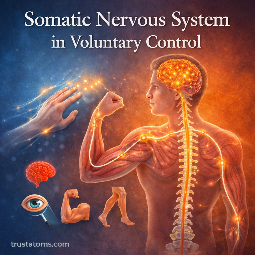
The somatic nervous system is responsible for voluntary control of skeletal muscles, allowing humans to consciously direct body movements. It also plays a major role in transmitting sensory information from the environment to the brain.
Understanding how the somatic nervous system functions helps explain how the brain coordinates movement, maintains posture, and interacts with the physical world.
What Is the Somatic Nervous System?
The somatic nervous system (SNS) is a division of the peripheral nervous system (PNS). It connects the central nervous system—which includes the brain and spinal cord—to skeletal muscles and sensory receptors throughout the body.
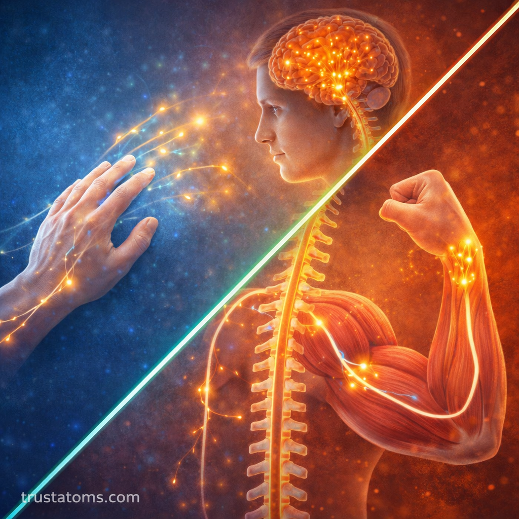
The system performs two primary functions:
- Carrying sensory signals from the body to the brain and spinal cord
- Sending motor commands from the central nervous system to skeletal muscles
Because of this two-way communication, the somatic nervous system enables both perception and action.
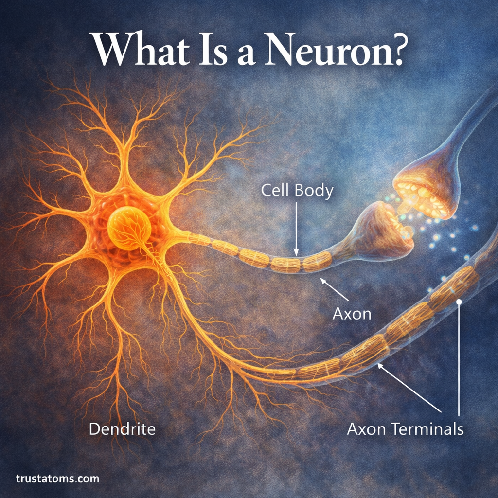
Key components include:
- Sensory neurons
- Motor neurons
- Skeletal muscles
- Sensory receptors in skin, muscles, and joints
Together, these components allow the brain to monitor body position and produce coordinated voluntary movements.
How Voluntary Movement Occurs
Voluntary movement is a complex process that involves several stages of neural communication between the brain, spinal cord, and muscles.
1. Decision and Motor Planning
Movement typically begins in areas of the brain responsible for planning and decision-making, such as the motor cortex.
During this stage:
- The brain determines the desired action
- Motor signals are organized and prepared
- Neural pathways are activated to initiate movement
For example, deciding to reach for an object activates motor circuits that control the arm and hand.
2. Signal Transmission Through the Spinal Cord
Once the brain generates a motor command, the signal travels through descending pathways in the spinal cord.
Motor neurons carry these signals from the spinal cord to skeletal muscles. These neurons act as the final pathway between the nervous system and the muscle fibers responsible for movement.
This stage ensures that the correct muscles receive the appropriate activation signals.
3. Muscle Activation
When motor neurons reach the muscle, they communicate with muscle fibers at specialized junctions called neuromuscular junctions.
At this connection point:
- Motor neurons release the neurotransmitter acetylcholine
- Muscle fibers receive the signal
- Muscle contraction begins
The contraction of muscle fibers produces the movement directed by the brain.
Sensory Feedback and Movement Control
Voluntary movement requires more than just motor commands. The body constantly sends sensory feedback to the brain to refine and adjust movement.
Sensory receptors in muscles, joints, and skin detect information such as:
- Body position
- Muscle stretch
- Pressure and touch
- Temperature and pain
This information travels back to the brain through sensory neurons.
The brain uses this feedback to:
- Correct movement errors
- Adjust force and direction
- Maintain balance and coordination
Without sensory feedback, voluntary movement would become inaccurate and difficult to control.
Structure of Somatic Neural Pathways
The somatic nervous system relies on specialized neural pathways that connect the brain to skeletal muscles.
These pathways typically involve two main neuron types.
Sensory (Afferent) Neurons
Sensory neurons transmit information from the body to the central nervous system.
They carry signals from:
- Skin receptors
- Muscle stretch receptors
- Joint sensors
- Pain and temperature receptors
These signals allow the brain to interpret environmental stimuli and body position.
Motor (Efferent) Neurons
Motor neurons transmit signals from the central nervous system to skeletal muscles.
Their functions include:
- Initiating muscle contraction
- Controlling movement strength
- Coordinating multiple muscle groups
Unlike the autonomic nervous system, which often uses multiple neuron connections, somatic motor pathways typically involve a single neuron directly connecting the spinal cord to the muscle.
The Role of the Neuromuscular Junction
The neuromuscular junction is the communication point between motor neurons and muscle fibers.
At this junction:
- A motor neuron releases acetylcholine.
- The neurotransmitter binds to receptors on the muscle fiber.
- Electrical activity spreads across the muscle cell membrane.
- Muscle contraction begins.
This mechanism allows neural signals to be converted into physical movement.
Because each motor neuron controls multiple muscle fibers, groups of fibers called motor units work together to produce smooth and coordinated actions.
Coordination with Other Nervous System Components
Although the somatic nervous system controls voluntary movement, it works closely with other neural systems to ensure precise coordination.
Important partners include:
The Cerebellum
The cerebellum helps refine movement by:
- Coordinating timing and accuracy
- Adjusting posture and balance
- Learning motor skills through repetition
The Basal Ganglia
The basal ganglia contribute to:
- Initiating movement patterns
- Regulating motor intensity
- Preventing unwanted movements
Spinal Reflex Circuits
Some somatic responses occur through spinal reflexes, which allow rapid reactions without requiring conscious processing.
Examples include:
- The knee-jerk reflex
- Withdrawal from painful stimuli
These reflexes help protect the body and maintain stability.
Importance of the Somatic Nervous System
The somatic nervous system allows humans to interact with their environment through intentional movement.
Key functions include:
- Voluntary muscle control
- Coordination of complex movements
- Sensory perception of the external environment
- Maintenance of posture and balance
Activities such as walking, writing, speaking, and athletic performance all rely on efficient somatic nervous system signaling.
Somatic Nervous System and Health
Damage to somatic neural pathways can disrupt voluntary movement and sensory perception.
Possible effects include:
- Muscle weakness or paralysis
- Loss of coordination
- Reduced reflex responses
- Impaired sensation
Conditions affecting somatic pathways may involve injuries to nerves, the spinal cord, or motor regions of the brain.
Research in neuroscience continues to explore how neural circuits controlling voluntary movement can be repaired or restored after injury.
Final Thoughts
The somatic nervous system is the primary pathway through which the brain controls voluntary movement. By linking sensory input with motor output, this system allows humans to respond to the environment with precise and coordinated actions.
From simple gestures to complex motor skills, the somatic nervous system enables conscious control of skeletal muscles while continuously adjusting movement through sensory feedback. Its intricate communication between the brain, spinal cord, and muscles forms the foundation of everyday human motion.