CNS Protection: Meninges and Cerebrospinal Fluid

Illustration showing the brain and spinal cord protected by the meninges and surrounded by cerebrospinal fluid.
Diagram showing how the meninges and cerebrospinal fluid protect the brain and spinal cord. trustatoms.com

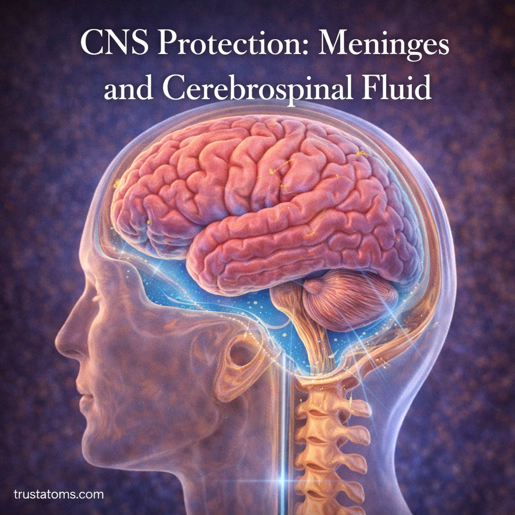
The central nervous system (CNS), which includes the brain and spinal cord, is one of the most vital and sensitive systems in the human body. Because of its importance, it requires multiple layers of protection to prevent damage from physical impact, infection, and environmental changes.

Two key protective components are the meninges and cerebrospinal fluid (CSF). Together, they create a supportive environment that cushions, stabilizes, and nourishes the brain and spinal cord.

Understanding how these structures work helps explain how the body protects its most critical control center.

Continue reading “CNS Protection: Meninges and Cerebrospinal Fluid”

Peripheral Nerves and Signal Routing

Illustration showing peripheral nerves branching from the spinal cord throughout the body to transmit signals between the brain and muscles.
Diagram showing peripheral nerves routing signals between the spinal cord and the rest of the body. trustatoms.com

Peripheral nerves form an extensive communication network that connects the brain and spinal cord to the rest of the body. These nerves carry signals that control movement, relay sensory information, and regulate many automatic body functions.

Through this network, the nervous system can quickly route messages between the central nervous system and muscles, organs, and sensory receptors. Without peripheral nerves, the brain would have no way to receive information from the body or send commands that control movement.

Understanding how peripheral nerves route signals provides insight into how the body coordinates sensation, motion, and internal regulation.

Continue reading “Peripheral Nerves and Signal Routing”

Cranial Nerves and Their Functions

Illustration showing cranial nerves extending from the brain to sensory organs including the eyes, ears, nose, tongue, and throat.
Medical illustration showing cranial nerves connecting the brain to sensory organs and muscles in the head and neck. trustatoms.com

Cranial nerves are a group of 12 pairs of nerves that originate directly from the brain rather than the spinal cord. These nerves play a critical role in controlling sensory perception, muscle movement, and essential body functions in the head, neck, and some internal organs.

They allow the brain to process sensory information such as smell, vision, hearing, taste, and facial sensation while also controlling movements like eye motion, facial expressions, and swallowing.

Understanding cranial nerves helps explain how the nervous system manages many everyday actions that occur without conscious thought.

Continue reading “Cranial Nerves and Their Functions”

Neural Integration in the Spinal Cord

Split diagram showing a reflex arc pulling a hand away from heat and neural pathways communicating between the spinal cord and brain.
Illustration showing a spinal reflex arc and communication between the spinal cord and brain during rapid protective responses. trustatoms.com

The spinal cord is more than just a pathway that carries signals between the brain and the body. It also acts as an important processing center that integrates sensory information and coordinates motor responses.

Neural integration in the spinal cord allows the body to respond quickly to stimuli, maintain posture, and coordinate movement without always needing direct involvement from the brain. This ability is especially important for reflexes and rapid protective responses.

Understanding how the spinal cord processes and integrates neural signals helps explain how the nervous system manages both automatic and voluntary actions.

Continue reading “Neural Integration in the Spinal Cord”

Proprioception and Body Position Sensing

Illustration showing how proprioception allows the brain to sense body position through signals from muscles, joints, and the nervous system.
Illustration explaining proprioception and how the body senses position and movement. trustatoms.com

Proprioception is the body’s ability to sense its own position, movement, and balance without needing to look. It allows you to walk without watching your feet, reach for objects accurately, and maintain posture automatically.

Often called the “sixth sense,” proprioception works through a network of sensory receptors in muscles, joints, and tendons that constantly send information to the brain about body position and movement.

This system operates continuously and mostly unconsciously, helping the brain coordinate movement, maintain balance, and prevent injury.

Continue reading “Proprioception and Body Position Sensing”

Pain Pathways and Nociception

Illustration showing pain pathways where nociceptors detect injury and transmit signals through the spinal cord to the brain.
Illustration depicting how nociceptors detect harmful stimuli and send pain signals through the spinal cord to the brain. trustatoms.com.

Pain is one of the body’s most important protective signals. When tissues are injured or exposed to potentially harmful conditions, the nervous system activates specialized pathways that alert the brain to danger. This process helps the body avoid further injury and initiate protective responses.

The biological detection and processing of harmful stimuli is known as nociception. Pain pathways carry these signals from sensory receptors in the body to the brain, where they are interpreted as the sensation of pain.

Understanding how nociception and pain pathways work provides insight into how the body detects injury, protects itself, and regulates responses to harmful stimuli.

Continue reading “Pain Pathways and Nociception”

Sensory Receptors and Transduction Mechanisms

Illustration showing human sensory receptors detecting stimuli such as light, sound, touch, heat, and smell and converting them into neural signals.
Illustration of sensory receptors detecting environmental stimuli and converting them into neural signals through sensory transduction. trustatoms.com.

The human body constantly gathers information about its surroundings and internal conditions. Every sight, sound, touch, taste, and smell begins with specialized structures known as sensory receptors. These receptors detect physical or chemical stimuli and convert them into electrical signals that the nervous system can interpret.

This process of converting environmental signals into neural impulses is called sensory transduction. It forms the foundation of perception, allowing the brain to interpret the world and respond appropriately.

Understanding sensory receptors and transduction mechanisms reveals how the nervous system translates external stimuli into meaningful sensory experiences.

Continue reading “Sensory Receptors and Transduction Mechanisms”

Somatic Nervous System in Voluntary Control

Illustration showing the somatic nervous system controlling voluntary muscle movement through signals between the brain, spinal cord, and skeletal muscles.
Illustration of the somatic nervous system demonstrating how the brain and spinal cord control voluntary skeletal muscle movement. trustatoms.com.

The human body performs thousands of movements every day, from simple actions like picking up a cup to complex activities such as writing, speaking, or playing sports. These movements are controlled by a specialized part of the nervous system known as the somatic nervous system.

The somatic nervous system is responsible for voluntary control of skeletal muscles, allowing humans to consciously direct body movements. It also plays a major role in transmitting sensory information from the environment to the brain.

Understanding how the somatic nervous system functions helps explain how the brain coordinates movement, maintains posture, and interacts with the physical world.

Continue reading “Somatic Nervous System in Voluntary Control”

Neurotransmitter Systems in Signal Modulation

Illustration of neurons communicating across a synapse representing neurotransmitter systems involved in signal modulation in the human brain.
Illustration showing how neurotransmitter systems modulate neural signals in the human brain. trustatoms.com.

The human nervous system relies on precise communication between billions of neurons. While electrical signals travel along nerve cells, the actual communication between neurons happens chemically through substances called neurotransmitters. These chemical messengers allow the brain and nervous system to regulate everything from movement and mood to memory and attention.

Neurotransmitter systems play a crucial role in signal modulation, meaning they influence how strongly or weakly signals are transmitted across neural networks. Rather than simply turning signals “on” or “off,” neurotransmitters fine-tune communication throughout the brain.

Understanding how these systems work helps explain many aspects of human behavior, cognition, and neurological health.

Continue reading “Neurotransmitter Systems in Signal Modulation”

Glial Cells in Neural Support

Illustration of glial cells supporting neurons in the nervous system, including astrocytes, oligodendrocytes, and microglia around neural networks.
Illustration showing glial cells supporting neurons by providing protection, nutrient delivery, and myelin formation in the nervous system. trustatoms.com

When people think about the nervous system, they often focus on neurons—the cells responsible for sending electrical signals throughout the brain and body. However, neurons do not function alone. The nervous system also relies on a large population of supportive cells known as glial cells.

Glial cells play essential roles in maintaining the health, structure, and efficiency of neural networks. They protect neurons, regulate the surrounding environment, provide nutrients, and assist in signal transmission.

Understanding glial cells reveals how the nervous system operates as a cooperative system rather than a collection of isolated neurons.

Continue reading “Glial Cells in Neural Support”