The female reproductive system is a complex and highly coordinated group of organs responsible for producing eggs, supporting fertilization, and nurturing the development of a fetus. It also plays a key role in hormone production, regulating processes such as the menstrual cycle and pregnancy.
Understanding female reproductive anatomy provides insight into fertility, hormonal health, and how the body prepares for and supports reproduction.
Primary Functions of the Female Reproductive System
The female reproductive system performs several essential functions:
- Egg production (oogenesis) – developing and releasing eggs (ova)
- Fertilization support – providing an environment where sperm can meet the egg
- Pregnancy support – nurturing and protecting a developing embryo and fetus
- Hormone regulation – producing hormones like estrogen and progesterone
Each organ contributes to one or more of these roles.
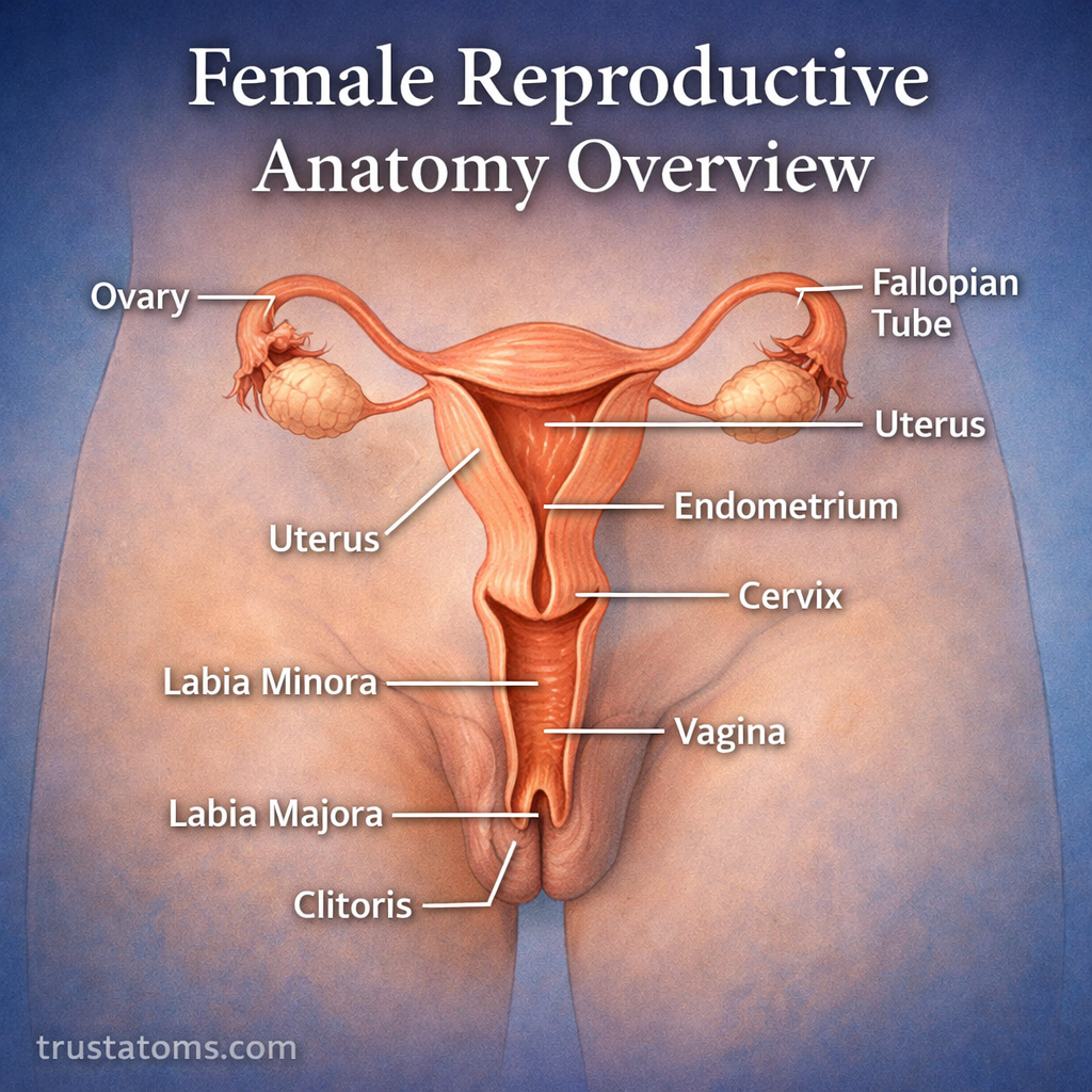
Key Structures of Female Reproductive Anatomy
External Organs (Vulva)
The external structures are collectively known as the vulva.
Labia Majora and Labia Minora
- Protect the internal reproductive organs
- Help maintain moisture and reduce friction
Clitoris
- A highly sensitive organ involved in sexual arousal
- Contains numerous nerve endings
Vaginal Opening
- Entrance to the vaginal canal
- Allows passage for menstrual flow, intercourse, and childbirth
Internal Organs
Vagina
The vagina is a muscular, flexible canal connecting the external body to the cervix.
- Serves as the passage for menstrual blood
- Receives sperm during intercourse
- Expands during childbirth
Cervix
The cervix is the lower, narrow part of the uterus.
- Connects the uterus to the vagina
- Produces cervical mucus that changes throughout the menstrual cycle
- Dilates during childbirth
Uterus
The uterus is a hollow, muscular organ where pregnancy develops.
- Lined with the endometrium, which thickens during the menstrual cycle
- Supports the growth of a fertilized egg
- Contracts during labor to deliver a baby
Fallopian Tubes
These tubes connect the ovaries to the uterus.
- Transport the egg from the ovary to the uterus
- Are the usual site of fertilization
Ovaries
The ovaries are the primary reproductive glands.
- Produce and release eggs (ovulation)
- Secrete hormones such as estrogen and progesterone
The Menstrual Cycle: How the System Works Together
The menstrual cycle is a repeating process that prepares the body for possible pregnancy. It typically lasts about 28 days, though variations are normal.
Main Phases of the Menstrual Cycle
- Menstrual Phase
- Shedding of the uterine lining
- Results in menstrual bleeding
- Follicular Phase
- Ovaries prepare an egg for release
- Estrogen levels rise
- Ovulation
- Release of a mature egg from the ovary
- Egg travels into the fallopian tube
- Luteal Phase
- Uterus prepares for possible implantation
- Progesterone levels increase
If fertilization does not occur, the cycle restarts.
Hormonal Control of Female Reproductive Anatomy
Hormones regulate nearly every function of the female reproductive system.
Key Hormones
- Estrogen – develops female characteristics and regulates the menstrual cycle
- Progesterone – prepares and maintains the uterus for pregnancy
- Follicle-stimulating hormone (FSH) – stimulates egg development
- Luteinizing hormone (LH) – triggers ovulation
These hormones are controlled by the brain, specifically the hypothalamus and pituitary gland.
Fertilization and Early Pregnancy
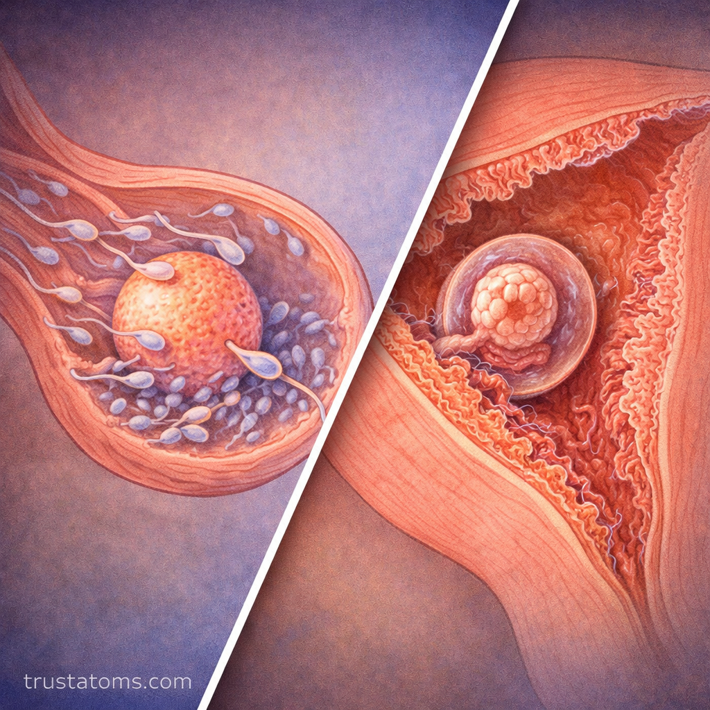
Fertilization typically occurs in the fallopian tubes when a sperm cell meets an egg.
- The fertilized egg (zygote) travels to the uterus
- It implants into the uterine lining
- The body begins producing hormones to support pregnancy
If implantation is successful, the menstrual cycle pauses during pregnancy.
Common Functions Working Together
The female reproductive system operates as an integrated system:
- Ovaries release eggs and hormones
- Fallopian tubes transport the egg
- Uterus prepares for implantation
- Cervix regulates entry and exit
- Vagina serves as the passageway
Each structure supports a different stage of reproduction.
Common Conditions Affecting Female Reproductive Anatomy
Several conditions can impact reproductive health:
- Endometriosis – growth of uterine-like tissue outside the uterus
- Polycystic ovary syndrome (PCOS) – hormonal imbalance affecting ovulation
- Fibroids – noncancerous growths in the uterus
- Cervical dysplasia – abnormal cell changes in the cervix
- Infertility – difficulty conceiving due to various factors
Understanding anatomy helps identify symptoms and seek appropriate care.
Why Understanding Female Reproductive Anatomy Matters
Knowledge of female reproductive anatomy is important for:
- Tracking menstrual health and fertility
- Understanding hormonal changes
- Supporting pregnancy and reproductive planning
- Recognizing early signs of potential health issues
It also empowers individuals to make informed healthcare decisions.
Final Thoughts
The female reproductive system is a highly organized and dynamic system that supports life from egg production to pregnancy. Each organ works in coordination with hormones to maintain reproductive health and function.
By understanding how these structures interact, it becomes easier to appreciate the complexity of the human body and the importance of maintaining overall reproductive wellness.