Lung volumes and capacities describe how much air your lungs can hold and how air moves during breathing. These measurements are essential for understanding respiratory health, diagnosing lung conditions, and evaluating overall lung function.
Whether you’re at rest or exercising, your lungs constantly adjust how much air they take in and release. Knowing how these volumes work gives insight into how efficiently your respiratory system performs.
What Are Lung Volumes and Capacities?
Lung volumes refer to specific amounts of air moved in and out of the lungs during breathing. Lung capacities are combinations of two or more lung volumes.
Together, they provide a complete picture of respiratory function.
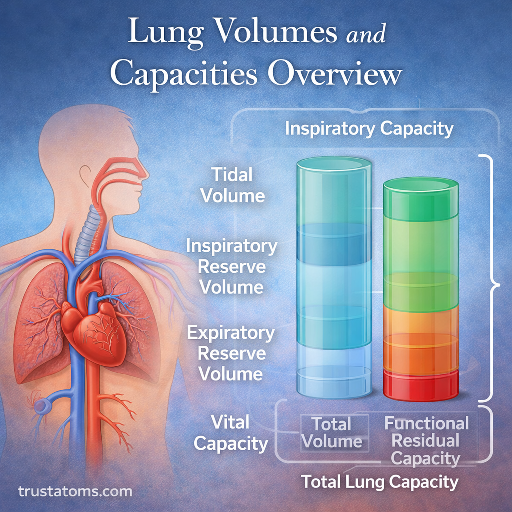
The Four Primary Lung Volumes
There are four main lung volumes that describe different phases of breathing.
1. Tidal Volume (TV)
Tidal volume is the amount of air you inhale or exhale during normal, relaxed breathing.
- Average: ~500 mL per breath
- Occurs during quiet breathing
- Requires minimal effort
This is the air you move in and out without thinking.
2. Inspiratory Reserve Volume (IRV)
Inspiratory reserve volume is the additional air you can inhale after a normal breath.
- Allows deeper inhalation when needed
- Used during exercise or exertion
- Expands lung capacity temporarily
3. Expiratory Reserve Volume (ERV)
Expiratory reserve volume is the extra air you can forcefully exhale after a normal breath.
- Helps clear more air from the lungs
- Important during activities like blowing out candles
- Engages abdominal and chest muscles
4. Residual Volume (RV)
Residual volume is the amount of air that remains in the lungs after a full exhalation.
- Prevents lung collapse
- Maintains continuous gas exchange
- Cannot be voluntarily expelled
Understanding Lung Capacities
Lung capacities are combinations of two or more lung volumes. They reflect how the lungs function as a whole.
1. Inspiratory Capacity (IC)
Inspiratory capacity is the total air you can inhale after a normal exhalation.
IC = Tidal Volume + Inspiratory Reserve Volume
- Represents maximum inhalation ability after resting breath
- Important for physical performance
2. Functional Residual Capacity (FRC)
Functional residual capacity is the air remaining in the lungs after a normal exhalation.
FRC = Expiratory Reserve Volume + Residual Volume
- Maintains steady gas exchange between breaths
- Acts as a buffer for oxygen and carbon dioxide levels
3. Vital Capacity (VC)
Vital capacity is the maximum amount of air you can exhale after a full inhalation.
VC = Tidal Volume + IRV + ERV
- Measures overall lung strength and flexibility
- Often used in pulmonary testing
4. Total Lung Capacity (TLC)
Total lung capacity is the maximum amount of air the lungs can hold.
TLC = Vital Capacity + Residual Volume
- Represents full lung expansion
- Varies based on body size, age, and fitness level
Why Lung Volumes and Capacities Matter
These measurements are essential for evaluating respiratory health and diagnosing conditions.
Key Uses Include:
- Assessing lung function in medical tests
- Diagnosing diseases like asthma or COPD
- Monitoring recovery or disease progression
- Evaluating athletic performance
Doctors often measure these values using a test called spirometry.
How Lung Volumes Are Measured
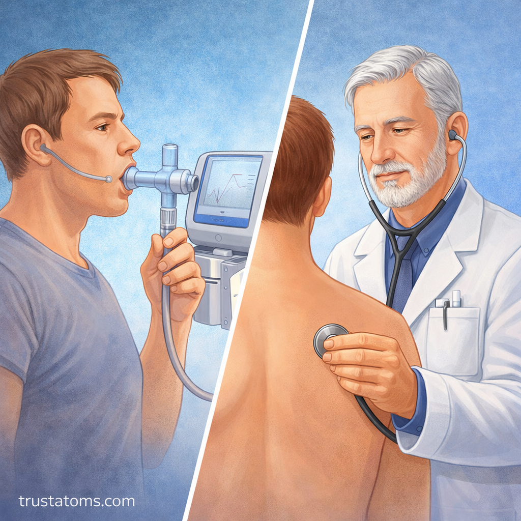
Lung volumes and capacities are typically measured using specialized equipment.
Common Methods:
- Spirometry: Measures airflow and volume during breathing
- Body plethysmography: Measures lung volume more precisely
- Gas dilution techniques: Estimate residual volume
Each method helps provide a clearer picture of lung health.
Factors That Affect Lung Volumes
Lung capacity is not the same for everyone. Several factors influence how much air your lungs can hold.
1. Age
- Lung capacity decreases over time
- Elasticity of lung tissue declines
2. Gender
- Males typically have larger lung volumes than females
3. Body Size
- Taller individuals generally have greater lung capacity
4. Physical Fitness
- Regular exercise can improve lung efficiency
- Athletes often have higher vital capacity
5. Health Conditions
- Diseases can restrict or obstruct airflow
- Examples include asthma, fibrosis, and emphysema
Common Lung Volume Disorders
Abnormal lung volumes can indicate respiratory problems.
Types of Disorders:
- Obstructive diseases: Difficulty exhaling (e.g., asthma, COPD)
- Restrictive diseases: Reduced lung expansion (e.g., fibrosis)
Effects on Lung Volumes:
- Obstructive: Increased residual volume, reduced airflow
- Restrictive: Reduced total lung capacity
Understanding these patterns helps guide diagnosis and treatment.
Lung Volumes in Everyday Life
You use different lung volumes throughout the day without realizing it.
Examples:
- Quiet breathing uses tidal volume
- Exercise increases inspiratory and expiratory reserves
- Deep breathing expands vital capacity
Your lungs continuously adapt to meet your body’s oxygen demands.
Final Thoughts
Lung volumes and capacities provide a detailed look at how your respiratory system functions. By measuring how much air moves in and out of the lungs, healthcare professionals can assess lung health, detect abnormalities, and guide treatment.
Understanding these concepts not only improves your knowledge of human anatomy but also highlights the importance of maintaining healthy lungs through lifestyle choices and proper care.