The spinal cord is a highly organized structure that connects the brain to the rest of the body. One of its most important features is its segmental organization—how it is divided into repeating sections, each linked to specific spinal nerve roots.
Spinal nerve roots act as communication highways, carrying sensory information into the spinal cord and motor commands out to muscles. Understanding how these roots are arranged helps explain everything from reflexes to patterns of pain and muscle weakness.
What Are Spinal Nerve Roots?
Spinal nerve roots are bundles of nerve fibers that emerge from the spinal cord. Each spinal nerve forms from two roots:
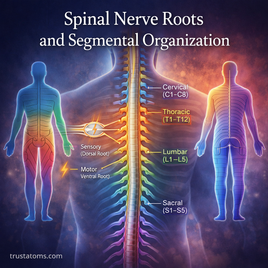
Dorsal (Posterior) Root
- Carries sensory information from the body to the spinal cord
- Includes signals for touch, pain, temperature, and position
- Contains the dorsal root ganglion (a cluster of sensory neuron cell bodies)
Ventral (Anterior) Root
- Carries motor commands from the spinal cord to muscles
- Controls voluntary movement and reflex actions
These two roots join together to form a spinal nerve, which then branches out to serve specific areas of the body.
Segmental Organization of the Spinal Cord
The spinal cord is divided into segments, each associated with a pair of spinal nerves. These segments are organized from top to bottom:
Major Spinal Regions
- Cervical (C1–C8): Neck, shoulders, arms, and diaphragm
- Thoracic (T1–T12): Chest and upper abdomen
- Lumbar (L1–L5): Lower back and legs
- Sacral (S1–S5): Pelvis, legs, and feet
- Coccygeal (Co1): Tailbone region
Each segment gives rise to a left and right spinal nerve, resulting in 31 pairs of spinal nerves.
How Spinal Nerves Are Formed
The process of spinal nerve formation follows a consistent pattern:
- Sensory signals enter through the dorsal root
- Motor signals exit through the ventral root
- The two roots merge to form a mixed spinal nerve
- The nerve branches into smaller pathways to reach target tissues
This organized structure allows efficient communication between the central nervous system and the body.
Dermatomes and Sensory Mapping
Dermatomes are areas of skin supplied by sensory fibers from a single spinal nerve root.
Why Dermatomes Matter
- Help map sensory loss in neurological exams
- Identify nerve root compression or injury
- Explain patterns of pain (e.g., radiating or “shooting” pain)
Examples
- C6 dermatome: Thumb and lateral forearm
- T4 dermatome: Area around the nipples
- L5 dermatome: Top of the foot and big toe
Because dermatomes follow a predictable pattern, they are widely used in clinical diagnosis.
Myotomes and Motor Control
Myotomes are groups of muscles controlled by motor fibers from a single spinal nerve root.
Key Functions
- Coordinate voluntary movement
- Help assess motor function during exams
- Indicate specific nerve root damage when weakness occurs
Examples
- C5: Shoulder abduction
- C7: Elbow extension
- L4: Knee extension
- S1: Ankle plantarflexion
Testing myotomes helps pinpoint where along the spinal cord or nerve pathway a problem may exist.
Reflex Arcs and Segmental Function
Reflexes are rapid, automatic responses that rely on spinal nerve roots and segmental organization.
How Reflex Arcs Work
- A sensory receptor detects a stimulus
- The signal travels through the dorsal root
- The spinal cord processes the information
- A motor response is sent through the ventral root
Common Reflexes
- Patellar reflex (L2–L4): Knee jerk
- Achilles reflex (S1–S2): Ankle jerk
- Biceps reflex (C5–C6): Arm flexion
Reflex testing is a key tool in neurological exams because it reveals how well specific spinal segments are functioning.
Clinical Significance of Spinal Nerve Roots
Spinal nerve roots are often involved in medical conditions, especially those affecting the spine.
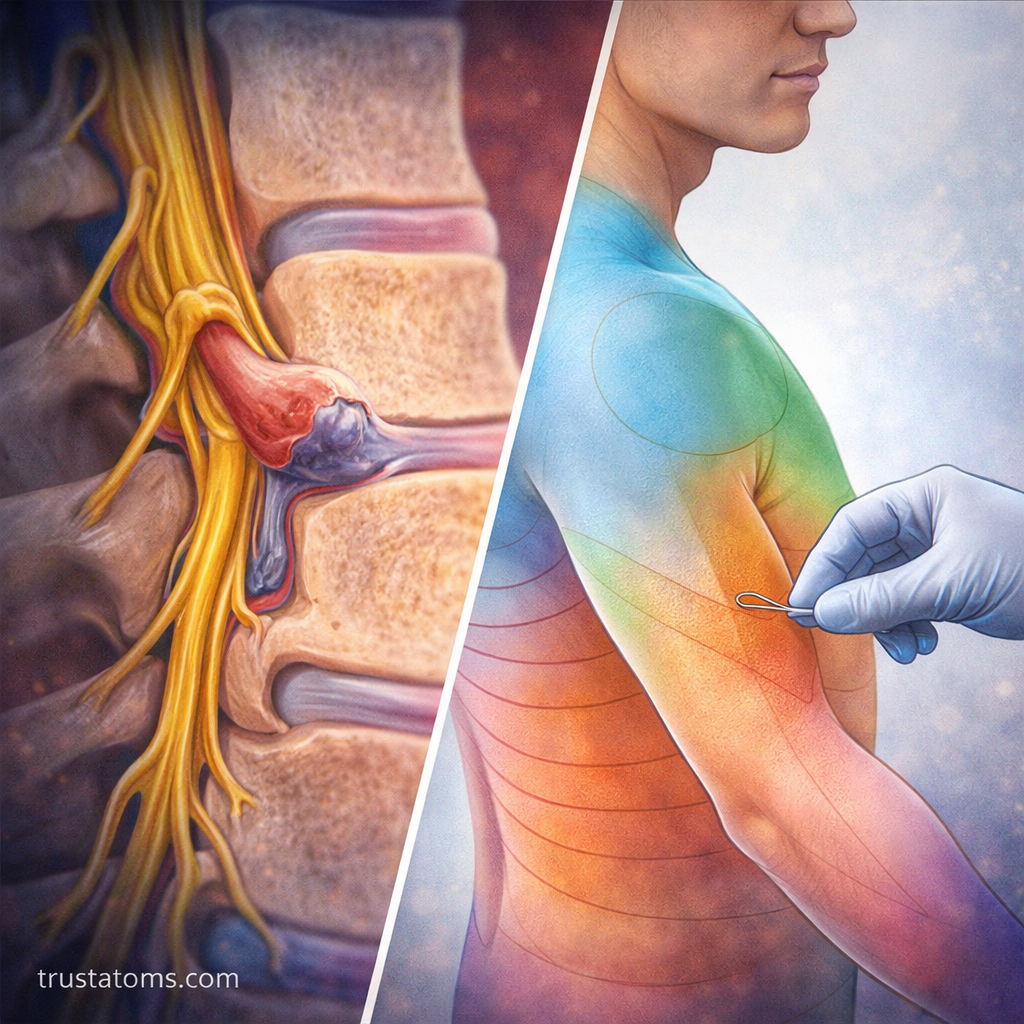
Nerve Root Compression (Radiculopathy)
Occurs when a spinal nerve root is compressed or irritated.
Common Causes
- Herniated discs
- Bone spurs (osteophytes)
- Spinal stenosis
Symptoms
- Radiating pain along a dermatome
- Numbness or tingling
- Muscle weakness in a myotome
Herniated Disc Example
When a disc bulges or ruptures, it can press on nearby nerve roots.
- Lumbar disc herniation often affects L4, L5, or S1 roots
- Symptoms may include sciatica (pain radiating down the leg)
Diagnostic Importance
Understanding segmental organization helps clinicians:
- Identify the level of spinal injury
- Localize neurological deficits
- Plan treatments such as physical therapy or surgery
Why Segmental Organization Matters
The spinal cord’s segmental design is not random—it provides a structured and efficient way to manage communication throughout the body.
Key Benefits
- Organized mapping of sensory and motor functions
- Faster reflex responses without brain involvement
- Easier diagnosis of neurological conditions
- Redundancy and overlap for functional resilience
This system ensures that even complex movements and sensations are coordinated smoothly.
Final Thoughts
Spinal nerve roots and segmental organization form the foundation of how the body communicates with the brain. By dividing the spinal cord into functional segments, the nervous system can efficiently manage movement, sensation, and reflexes.
From diagnosing nerve injuries to understanding patterns of pain, this structured design is essential for both everyday function and clinical practice. As research advances, deeper insights into spinal organization continue to improve treatments for neurological and musculoskeletal conditions.