Pleura and Pleural Pressure

Illustration of pleura and pleural pressure showing lung expansion within the chest cavity and pleural space.
Diagram of the pleural layers and pressure changes that keep the lungs expanded during breathing. trustatoms.com.

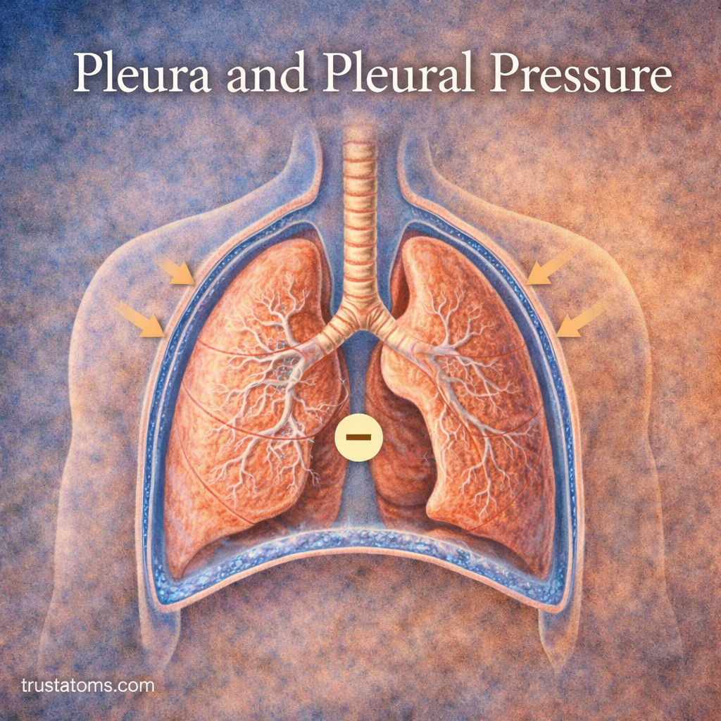
The lungs do not function in isolation—they rely on a thin, fluid-filled membrane system called the pleura to move smoothly within the chest cavity. Along with this structure, pleural pressure plays a crucial role in keeping the lungs expanded and enabling efficient breathing.

Understanding the pleura and pleural pressure helps explain how the lungs stay inflated and what happens when this system is disrupted.

Continue reading “Pleura and Pleural Pressure”

Respiratory Muscles Beyond the Diaphragm

Illustration of respiratory muscles including intercostals, neck muscles, and abdominal muscles supporting breathing beyond the diaphragm.
Diagram showing accessory respiratory muscles that assist breathing alongside the diaphragm. trustatoms.com.

When most people think about breathing, they think of the diaphragm—the primary muscle responsible for inhalation. However, breathing also relies on a group of additional muscles that assist in expanding and compressing the lungs, especially during physical activity or respiratory stress.

These supporting muscles, often called accessory respiratory muscles, play a crucial role in maintaining efficient airflow when the body’s demand for oxygen increases.

Continue reading “Respiratory Muscles Beyond the Diaphragm”

Ventilation–Perfusion Matching Principles

Illustration showing ventilation and perfusion matching in alveoli with comparison of normal and impaired airflow and blood flow.
Diagram of ventilation–perfusion matching showing how balanced airflow and blood flow optimize gas exchange in the lungs. trustatoms.com.

Ventilation–perfusion (V/Q) matching is a key principle of respiratory physiology that ensures oxygen enters the blood efficiently and carbon dioxide is removed effectively. It refers to the balance between airflow reaching the alveoli (ventilation) and blood flow reaching the lungs (perfusion).

When ventilation and perfusion are properly matched, gas exchange is optimized. When they are not, breathing becomes less efficient—even if the lungs themselves appear structurally normal.

Continue reading “Ventilation–Perfusion Matching Principles”

Partial Pressure and Gas Diffusion

Illustration showing partial pressure and gas diffusion between alveoli and capillaries with oxygen and carbon dioxide movement.
Diagram of gas exchange in the lungs illustrating how partial pressure drives oxygen into the blood and carbon dioxide out. trustatoms.com.

Gas exchange in the lungs depends on a fundamental principle of physics: gases move from areas of higher pressure to areas of lower pressure. In the respiratory system, this process is driven by differences in partial pressure, allowing oxygen to enter the blood and carbon dioxide to leave it.

Understanding partial pressure and gas diffusion is essential for explaining how breathing supports life at a cellular level.

Continue reading “Partial Pressure and Gas Diffusion”

Surfactant Function in Alveolar Stability

Illustration of surfactant function in alveoli showing reduced surface tension and stabilized air sacs in the lungs.
Close-up of alveoli demonstrating how surfactant reduces surface tension to keep air sacs open and stable. trustatoms.com.

Surfactant is a specialized substance in the lungs that plays a critical role in keeping the alveoli stable and functional. Without it, breathing would require significantly more effort, and the tiny air sacs responsible for gas exchange could collapse.

Understanding how surfactant works provides key insight into respiratory efficiency, lung development, and conditions that affect breathing.

Continue reading “Surfactant Function in Alveolar Stability”

Alveolar Structure and Surface Area

Illustration of alveoli structure and surface area showing air sacs and surrounding capillaries in the lungs.
Detailed view of alveoli highlighting their structure and large surface area for efficient gas exchange. trustatoms.com.

Alveoli are tiny air sacs in the lungs where oxygen and carbon dioxide are exchanged between the air and the bloodstream. Although each alveolus is microscopic, their combined structure creates a massive surface area that is essential for efficient respiration.

Understanding alveolar structure and surface area helps explain how the lungs meet the body’s oxygen demands—and why even small damage to this system can have major consequences.

Continue reading “Alveolar Structure and Surface Area”

Bronchioles and Airway Resistance

Illustration showing bronchioles and airway resistance with normal and constricted airways in the lungs.
Simple diagram of bronchioles demonstrating how airway narrowing increases resistance in the lungs. trustatoms.com.

Bronchioles are the smallest airways in the respiratory system, playing a critical role in regulating airflow to the lungs. While they are tiny compared to the trachea and bronchi, their structure and function have a major impact on airway resistance and overall breathing efficiency.

Understanding how bronchioles influence airflow helps explain normal respiration, exercise performance, and common respiratory conditions such as asthma.

Continue reading “Bronchioles and Airway Resistance”

Trachea and Bronchi in Airflow Distribution

Diagram showing airflow moving through the trachea into branching bronchi and lungs.
Air flows through the trachea and bronchi, distributing oxygen throughout the lungs. trustatoms.com.

The trachea and bronchi play a central role in directing air from the upper respiratory tract into the lungs. These structures act as a branching airway system, ensuring that inhaled air is efficiently distributed to every part of the lungs.

Understanding how airflow moves through the trachea and bronchi provides insight into how the respiratory system supports breathing, oxygen delivery, and overall lung function.

Continue reading “Trachea and Bronchi in Airflow Distribution”

Upper vs Lower Respiratory Tract

Diagram comparing the upper and lower respiratory tract including nasal passages, throat, trachea, and lungs.
The respiratory system is divided into upper and lower tracts, each with distinct roles in breathing and protection. trustatoms.com.

The respiratory system is divided into two main sections: the upper respiratory tract and the lower respiratory tract. Together, these structures work to move air in and out of the body, filter harmful particles, and enable gas exchange.

Understanding the differences between these two regions helps clarify how breathing works and how various respiratory conditions develop.

Continue reading “Upper vs Lower Respiratory Tract”

Respiratory Rate and Ventilation Control

Illustration showing brain control of breathing with lungs exchanging oxygen and carbon dioxide.
The brain regulates breathing rate to balance oxygen intake and carbon dioxide removal. trustatoms.com.

Breathing is something most people rarely think about, yet it is constantly adjusted to meet the body’s needs. Respiratory rate and ventilation control ensure that oxygen is delivered efficiently and carbon dioxide is removed at the right pace.

This system is highly responsive, automatically adapting to changes in activity, environment, and internal chemistry.

Continue reading “Respiratory Rate and Ventilation Control”