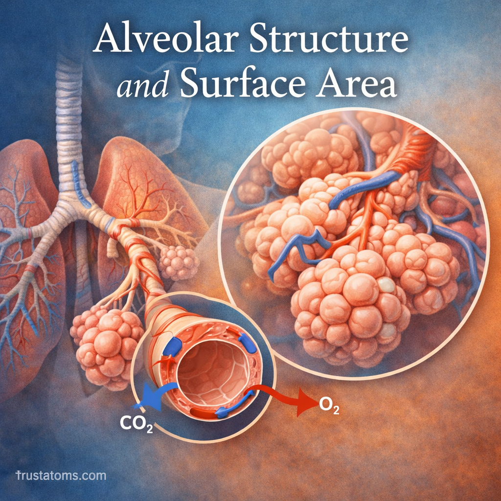
Alveoli are microscopic air sacs in the lungs whose thin walls and extensive combined surface area allow oxygen to enter the bloodstream and carbon dioxide to leave it. Their close contact with pulmonary capillaries creates a short diffusion distance, making gas exchange efficient enough to support the body’s continuous oxygen needs.
In human anatomy, alveoli demonstrate how microscopic structures support the function of an entire organ. Their number, shape, surface lining, and surrounding blood vessels all influence how effectively the lungs exchange gases.
What Are Alveoli?
Alveoli are tiny air-filled sacs in the gas-exchanging regions of the respiratory system, located beyond the terminal bronchioles. They are the primary sites of gas exchange.
Air reaches the alveoli through respiratory bronchioles, alveolar ducts, and alveolar sacs. Rather than existing as isolated balloons, neighboring alveoli share thin walls called interalveolar septa, which contain capillaries and supporting tissue.
Adult human lungs contain several hundred million alveoli, although estimates vary with lung size and measurement methods. Together, they provide an extensive surface for contact between air and blood.
The relationship between available surface area and the movement of gases is examined in diffusion capacity in lung function. The pressure differences that drive oxygen and carbon dioxide across the exchange barrier are explained in partial pressure and gas diffusion.
The Microscopic Structure of an Alveolus
Alveoli have specialized cells and supporting structures that keep the air spaces open while allowing gases to cross into and out of the blood.
Type I Alveolar Cells: The Thin Exchange Surface
Type I alveolar epithelial cells are extremely thin, flattened cells covering most of the alveolar surface. Their shape minimizes the distance oxygen and carbon dioxide must travel between alveolar air and nearby capillaries.
The gas-exchange barrier includes the alveolar lining, epithelial cells, a thin interstitial region with basement membranes, and capillary endothelial cells. In its thinnest regions, this barrier is less than one micrometer thick.
Type II Alveolar Cells: Producing Surfactant
Type II alveolar cells produce pulmonary surfactant, a mixture of lipids and proteins that reduces surface tension at the air-liquid interface. Lower surface tension helps stabilize alveoli and reduces the effort required to inflate the lungs.
These cells also contribute to alveolar repair by dividing and producing replacement epithelial cells following certain injuries.
Pulmonary Capillaries: Bringing Blood Close to Air
A dense capillary network runs through the thin walls separating neighboring alveoli. Blood arriving from the pulmonary arteries has relatively low oxygen partial pressure and higher carbon dioxide partial pressure than alveolar air under normal conditions.
As blood moves through these capillaries, oxygen enters it while carbon dioxide moves toward the alveolar spaces. Continuous circulation helps maintain the pressure differences needed for diffusion.
How Much Surface Area Do Human Alveoli Provide?
The total alveolar surface area in healthy adult lungs is commonly estimated at approximately 50–100 square meters, depending on lung inflation, body size, and measurement technique. This is a large exchange surface packed inside the chest.
Surface area matters because gas diffusion increases when more membrane is available for gases to cross, assuming other conditions remain comparable.
Three structural features make this possible:
- Numerous small air sacs: Hundreds of millions of alveoli collectively provide far more surface area than a few large chambers would.
- Shared alveolar walls: Closely packed air spaces allow an extensive exchange surface within a relatively compact volume.
- Extensive capillary contact: Blood vessels closely follow the alveolar surfaces, making much of the available area useful for gas exchange.
A Simple Example: Why Many Small Sacs Provide More Area
Imagine two spherical air spaces containing the same total volume of air. One is a single sphere, while the other consists of eight smaller spheres, each with half the radius of the original sphere.
Because the volume of a sphere depends on the cube of its radius, the eight smaller spheres together contain the same total volume as the larger sphere. However, their combined surface area is twice as large.
Real alveoli are not perfect spheres, and their geometry changes during breathing. Still, this mathematical example illustrates why dividing a volume into numerous small compartments can greatly increase the surface available for gas exchange.
How Oxygen and Carbon Dioxide Cross the Alveolar Wall
Gas exchange occurs through passive diffusion, driven by differences in the partial pressures of oxygen and carbon dioxide.
- Fresh air enters the gas-exchanging regions during inhalation.
- Oxygen diffuses from alveolar air across the alveolar-capillary membrane into pulmonary blood.
- Most oxygen binds to hemoglobin inside red blood cells for transport through the circulation.
- Carbon dioxide diffuses from pulmonary blood into alveolar air.
- Ventilation removes carbon dioxide from the lungs during exhalation.
The process follows principles summarized by Fick’s law of diffusion: greater surface area and larger partial-pressure differences favor gas transfer, while a thicker diffusion barrier slows it. The gas’s physical properties also influence its diffusion rate.
Factors That Affect Alveolar Efficiency
Available Surface Area
When alveolar walls are destroyed, the available exchange surface decreases. Even if air continues reaching the lungs, less functional membrane remains for oxygen and carbon dioxide transfer.
Thickness of the Exchange Barrier
Inflammation, scarring, or fluid accumulation can increase the distance gases must cross. Oxygen transfer is particularly vulnerable when diffusion becomes limited.
Ventilation and Blood Flow
Effective gas exchange requires both air reaching the alveoli and blood flowing through nearby capillaries. A mismatch between ventilation and perfusion can reduce oxygen uptake even when alveolar walls remain structurally intact.
Surfactant and Alveolar Stability
Surfactant reduces surface tension and helps prevent alveolar collapse, especially as lung volume decreases during exhalation. Without sufficient surfactant, breathing becomes more difficult and some air spaces may collapse. The mechanisms are described further in surfactant function in alveolar stability.
How Lung Diseases Damage Alveolar Structure
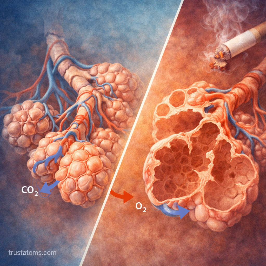
Different respiratory diseases interfere with alveolar function through distinct mechanisms. Some destroy the exchange surface, while others thicken the barrier or fill air spaces with fluid.
Emphysema: Loss of Alveolar Walls
Emphysema damages and destroys alveolar walls, producing enlarged air spaces with less total surface area. Loss of elastic recoil also makes exhalation more difficult and contributes to air trapping.
Pulmonary Edema: Fluid Interfering With Gas Exchange
Pulmonary edema occurs when excess fluid accumulates in lung tissue and potentially inside alveoli. This can interfere with ventilation, increase diffusion distance, and reduce blood oxygen levels.
Pulmonary Fibrosis and Acute Lung Injury
Pulmonary fibrosis causes scarring that can thicken and stiffen lung tissue, reducing diffusion efficiency and making lung expansion harder. Acute respiratory distress syndrome can damage the alveolar-capillary barrier, causing inflammation, fluid leakage, and impaired oxygenation.
Frequently Asked Questions
How many alveoli are in the human lungs?
Adult human lungs typically contain several hundred million alveoli in total. The exact number varies among individuals and depends on the methods used to estimate it.
Why are alveolar walls so thin?
Thin alveolar walls minimize the distance oxygen and carbon dioxide must cross during diffusion. Their close association with capillaries allows gases to move efficiently between air and blood.
Can damaged alveoli regenerate?
Type II alveolar cells can help repair certain injuries by replacing damaged epithelial cells. However, extensive destruction of alveolar walls, as occurs in emphysema, is generally not fully reversible, and normal gas-exchange surface area may not be restored.
Alveolar efficiency depends on more than the number of air sacs: thin exchange barriers, extensive surface area, surfactant, ventilation, and capillary blood flow must work together. Diseases that destroy alveolar walls or disrupt their surrounding tissues demonstrate how changes at the microscopic level can substantially affect breathing and blood oxygenation.
This article is for general education and is not medical advice. Talk to a qualified health professional about any medical concern.
Sources: West’s Respiratory Physiology: The Essentials; Guyton and Hall Textbook of Medical Physiology; National Heart, Lung, and Blood Institute.