Movement is one of the most essential abilities of the human body. From walking and breathing to writing and speaking, nearly every voluntary action depends on specialized nerve cells called motor neurons. These cells transmit signals from the brain and spinal cord to muscles, allowing them to contract and produce movement.
Motor neurons form a crucial part of the nervous system’s communication network. They convert electrical signals generated in the central nervous system into muscle activity, coordinating everything from simple reflexes to complex athletic movements.
Understanding how motor neurons work helps explain how the body controls movement, maintains posture, and adapts to changing physical demands.
What Are Motor Neurons?
Motor neurons are specialized nerve cells that carry signals from the central nervous system (CNS) to muscles or glands. Their main function is to initiate muscle contraction.
They act as messengers between the brain or spinal cord and the muscles responsible for movement.
Key characteristics of motor neurons include:
- Long nerve fibers (axons) that extend from the spinal cord to muscles
- Ability to transmit electrical impulses rapidly
- Connections with muscle fibers at specialized junctions
- Integration with sensory and control circuits in the nervous system
Without motor neurons, muscles would not receive the instructions needed to move.
The Two Main Types of Motor Neurons
Movement control depends on two primary categories of motor neurons working together.
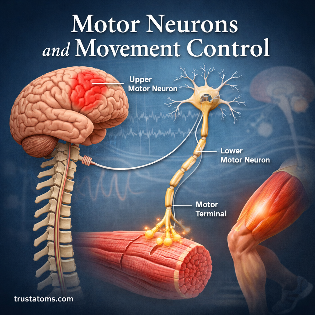
Upper Motor Neurons
Upper motor neurons originate in the brain, specifically in the motor cortex. Their job is to send movement commands down to the spinal cord.
Functions of upper motor neurons include:
- Initiating voluntary movement
- Controlling muscle tone
- Regulating posture and coordination
- Relaying signals to lower motor neurons
These neurons do not directly contact muscles. Instead, they communicate with lower motor neurons located in the spinal cord.
Lower Motor Neurons
Lower motor neurons carry signals from the spinal cord to the muscles themselves.
Their roles include:
- Directly activating muscle fibers
- Producing muscle contractions
- Controlling strength and precision of movements
- Maintaining muscle tone
Damage to lower motor neurons can lead to muscle weakness or paralysis because the muscles lose their direct neural input.
Structure of a Motor Neuron
Like other neurons, motor neurons have specialized structures that allow them to transmit information.
The main components include:
Cell Body (Soma)
The cell body contains the nucleus and essential cellular machinery responsible for maintaining the neuron.
Inside the nucleus, genetic information stored within chromosomes helps regulate the proteins required for neuron function and communication. In eukaryotic cells, DNA is packaged into chromosomes within the nucleus where it controls cellular activity and gene expression.
Dendrites
Dendrites are short branches that receive signals from other neurons.
They collect incoming information and send it toward the cell body.
Axon
The axon is a long extension that carries electrical impulses away from the cell body.
Motor neuron axons can be extremely long—sometimes extending from the spinal cord to muscles in the feet.
Axon Terminals
At the end of the axon are terminals that connect to muscle fibers through structures called neuromuscular junctions.
These junctions allow signals to pass from the neuron to the muscle.
The Neuromuscular Junction
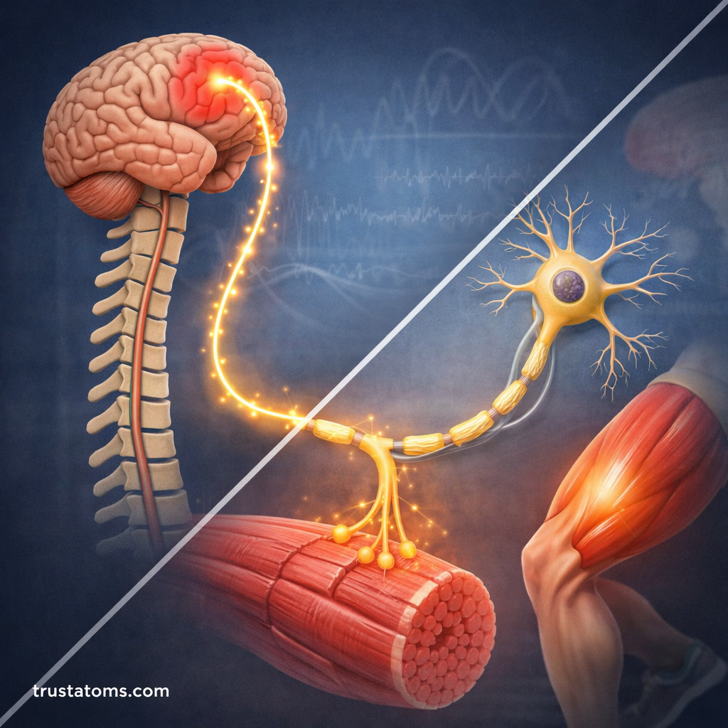
The neuromuscular junction (NMJ) is the point where a motor neuron communicates with a muscle fiber.
This connection is essential for movement.
The process works as follows:
- A motor neuron sends an electrical signal down its axon.
- The signal reaches the neuromuscular junction.
- The neuron releases a neurotransmitter called acetylcholine.
- Acetylcholine binds to receptors on the muscle cell.
- The muscle fiber contracts.
This rapid communication happens in milliseconds and allows for precise movement control.
How the Brain Controls Movement
Movement begins in the brain.
Several regions work together to plan, initiate, and coordinate motor activity.
Motor Cortex
Located in the frontal lobe of the brain, the motor cortex is responsible for initiating voluntary movements.
Different regions of the motor cortex control different body parts.
Cerebellum
The cerebellum plays a major role in coordination, balance, and timing of movements.
It helps refine motor commands and correct errors in motion.
Basal Ganglia
The basal ganglia regulate movement intensity and smoothness.
They help prevent unwanted movements and ensure actions are performed smoothly.
Spinal Cord
The spinal cord acts as a relay center between the brain and muscles.
It also manages reflexes that allow rapid responses without needing direct brain involvement.
Motor Units and Muscle Control
A motor unit consists of:
- One motor neuron
- All the muscle fibers it controls
Motor units allow muscles to produce varying levels of force.
For example:
Small motor units control fine movements such as:
- Eye movement
- Finger control
- Facial expressions
Large motor units control powerful movements such as:
- Leg muscles used in running
- Back muscles used in lifting
The nervous system adjusts force by activating different numbers of motor units.
Reflexes and Automatic Movement
Not all movements require conscious control.
Reflexes are automatic responses that help protect the body.
Reflex pathways often involve:
- A sensory neuron detecting a stimulus
- The spinal cord processing the signal
- A motor neuron activating a muscle response
Examples of reflexes include:
- Pulling your hand away from a hot surface
- Knee-jerk reflex during medical exams
- Rapid balance corrections while walking
These responses occur quickly because they bypass slower brain processing.
The Role of Motor Neurons in Posture and Balance
Motor neurons also maintain posture and stability even when you are not consciously moving.
They constantly adjust muscle activity to:
- Keep the body upright
- Stabilize joints
- Maintain muscle tone
- Coordinate balance
The nervous system continuously sends small signals to muscles to keep them slightly contracted, preventing collapse under gravity.
Motor Neuron Disorders
Damage to motor neurons can significantly affect movement.
Several neurological conditions involve degeneration of motor neurons.
Amyotrophic Lateral Sclerosis (ALS)
ALS causes progressive loss of motor neurons in the brain and spinal cord.
Symptoms often include:
- Muscle weakness
- Difficulty speaking or swallowing
- Loss of voluntary movement
Spinal Muscular Atrophy (SMA)
SMA affects lower motor neurons, leading to muscle weakness and reduced muscle mass.
Multiple Sclerosis (MS)
In MS, damage to the protective myelin sheath around nerve fibers disrupts signal transmission, affecting movement and coordination.
Research continues to explore treatments that protect or restore motor neuron function.
How Motor Neurons Enable Complex Movement
Human movement requires remarkable coordination between the nervous system and muscles.
Motor neurons allow this by:
- Translating brain signals into muscle contractions
- Coordinating multiple muscle groups simultaneously
- Adjusting force and precision in real time
- Integrating feedback from sensory neurons
For example, walking involves:
- Signals from the motor cortex
- Balance adjustments from the cerebellum
- Reflex feedback from the spinal cord
- Activation of dozens of muscles through motor neurons
All of these processes occur seamlessly, allowing smooth and controlled motion.
Why Motor Neurons Are Essential for Life
Motor neurons are responsible for nearly every physical action the body performs.
They control:
- Walking and running
- Breathing
- Speaking
- Facial expressions
- Hand coordination
- Posture and balance
Even subtle movements like blinking or swallowing depend on motor neuron activity.
Without these cells, the brain would not be able to communicate with the body’s muscles.
Final Thoughts
Motor neurons are the critical link between the nervous system and the muscular system. By transmitting signals from the brain and spinal cord to muscles, they make voluntary movement, posture, reflexes, and coordination possible.
Through intricate networks involving the motor cortex, spinal cord, and neuromuscular junctions, motor neurons allow humans to perform both delicate tasks and powerful movements. Their health and proper function are essential for everyday life, and ongoing research continues to deepen our understanding of how these remarkable cells control movement.