Fetal circulation is a specialized system that allows a developing baby to receive oxygen and nutrients from the mother while its own lungs are not yet functioning. Unlike postnatal circulation, fetal blood flow uses unique structures to bypass the lungs and liver, ensuring efficient delivery of oxygen-rich blood.
Understanding fetal circulation provides insight into how the body adapts before birth—and how it transitions immediately after delivery.
What Is Fetal Circulation?
Fetal circulation refers to the pattern of blood flow in a fetus during pregnancy. Since the fetus does not breathe air, oxygen is supplied through the placenta instead of the lungs.
Key Differences from Adult Circulation
- Oxygen comes from the placenta, not the lungs
- Blood bypasses the lungs and partially bypasses the liver
- Special shunts redirect blood flow efficiently
These adaptations allow the fetus to thrive in the womb.
Role of the Placenta
The placenta acts as the fetus’s lifeline.
Main Functions
- Supplies oxygen from the mother’s blood
- Delivers nutrients (glucose, amino acids, etc.)
- Removes carbon dioxide and waste products
Blood does not mix directly between mother and fetus, but exchange occurs across a thin membrane.
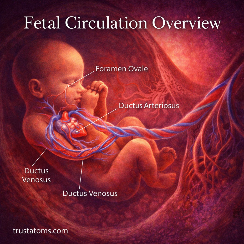
Key Structures in Fetal Circulation
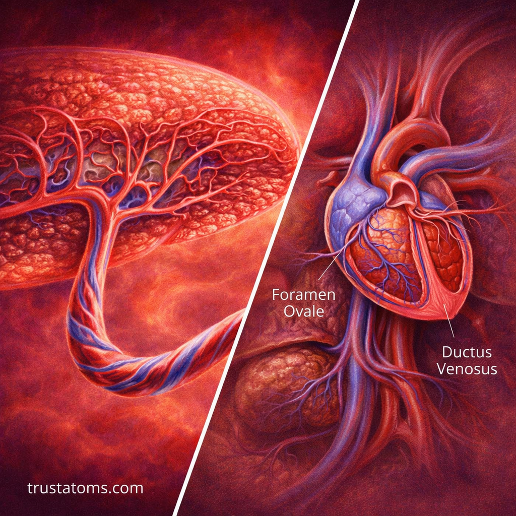
Fetal circulation relies on three important shunts that redirect blood flow.
1. Ductus Venosus
- Connects the umbilical vein to the inferior vena cava
- Allows oxygenated blood to bypass most of the liver
- Ensures rapid delivery to the heart
2. Foramen Ovale
- Opening between the right and left atria
- Allows blood to bypass the lungs
- Directs oxygen-rich blood toward the brain and heart
3. Ductus Arteriosus
- Connects the pulmonary artery to the aorta
- Diverts blood away from the lungs
- Sends blood directly into systemic circulation
These structures are essential for efficient fetal blood flow.
Step-by-Step Blood Flow in the Fetus
Here’s how blood moves through the fetal circulatory system:
- Oxygenated blood enters through the umbilical vein
- Blood passes through the ductus venosus, bypassing the liver
- Blood enters the inferior vena cava and reaches the right atrium
- Most blood flows through the foramen ovale into the left atrium
- Blood moves into the left ventricle and is pumped into the aorta
- Some blood goes to the right ventricle and into the pulmonary artery
- Blood bypasses the lungs via the ductus arteriosus
- Deoxygenated blood returns to the placenta through umbilical arteries
This system ensures that oxygen-rich blood reaches vital organs efficiently.
Why the Lungs Are Bypassed
Before birth, the lungs are filled with fluid and not used for oxygen exchange.
Reasons for Bypass
- No air is present in the lungs
- Blood vessels in the lungs have high resistance
- Oxygen is supplied more efficiently through the placenta
By bypassing the lungs, the fetus conserves energy and maintains effective circulation.
Distribution of Oxygenated Blood
Not all tissues receive the same oxygen concentration.
Priority Organs
- Brain
- Heart
- Upper body
These organs receive the most oxygen-rich blood to support development.
Lower Priority Areas
- Lower body
- Some abdominal organs
This selective distribution supports growth where it is needed most.
Changes at Birth
At birth, the circulatory system undergoes rapid transformation.
What Triggers the Change
- First breath expands the lungs
- Blood flow to the lungs increases
- Placental circulation stops when the umbilical cord is cut
Closure of Fetal Shunts
- Foramen ovale closes due to pressure changes
- Ductus arteriosus constricts and eventually becomes a ligament
- Ductus venosus closes as liver circulation increases
These changes convert fetal circulation into the adult pattern.
Clinical Significance
Problems in fetal circulation can lead to medical conditions.
Common Issues
- Patent foramen ovale (PFO) – incomplete closure
- Patent ductus arteriosus (PDA) – persistent connection
- Congenital heart defects affecting blood flow
Why It Matters
- Early detection helps guide treatment
- Some conditions resolve naturally
- Others may require medical or surgical intervention
Understanding fetal circulation is essential in neonatal and pediatric care.
Comparison: Fetal vs Adult Circulation
Fetal Circulation
- Oxygen from placenta
- Blood bypasses lungs and partially bypasses liver
- Specialized shunts present
Adult Circulation
- Oxygen from lungs
- Full circulation through liver and lungs
- No shunts
This transition is one of the most dramatic physiological changes in human life.
Everyday Importance of Fetal Circulation
Although it functions only before birth, fetal circulation is critical for development.
Why It Matters
- Supports organ growth and brain development
- Ensures efficient oxygen delivery
- Prepares the body for life outside the womb
A properly functioning fetal circulation system is essential for a healthy start to life.
Final Thoughts
Fetal circulation is a remarkable adaptation that allows a developing baby to thrive without using its lungs. Through specialized structures like the ductus venosus, foramen ovale, and ductus arteriosus, blood is efficiently directed to the most important organs.
At birth, this system transforms rapidly into adult circulation, marking one of the most important transitions in human physiology.