Joints are connections between bones that provide stability, permit movement, or accomplish both functions. Their structure determines whether they remain nearly immovable, allow limited flexibility, or support a wide range of movements such as bending, rotating, and reaching.
Studying joints is a fundamental part of human anatomy because the skeleton must support body weight while allowing coordinated movement. Understanding joint types and their range of motion helps explain how the human body performs both simple and complex movements.
What Is a Joint?
A joint, also called an articulation, is a location where two or more bones meet. Some joints allow extensive movement, while others primarily provide stability and structural support.
Joints can be classified by the tissues connecting their bones or by the amount of movement they permit. Their function also depends on surrounding muscles, ligaments, tendons, and cartilage.
The shoulder demonstrates how joint shape influences movement. Shoulder Joint Mechanics and Mobility explains how its relatively shallow socket permits extensive arm motion.
However, greater mobility requires effective support from surrounding tissues. The interaction between these structures is explored in Joint Stability and Supporting Structures.
Three Structural Types of Joints
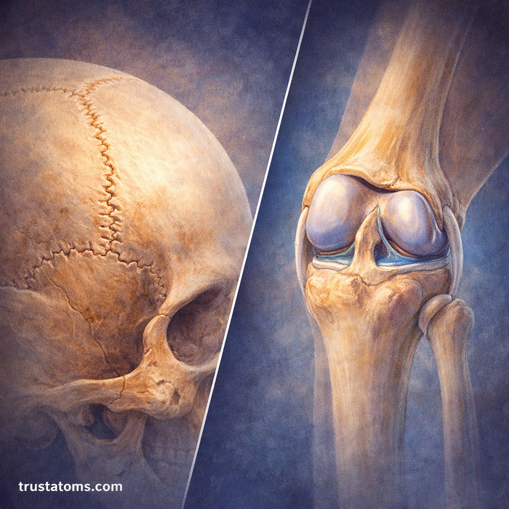
Anatomists divide joints into three structural categories based on how their bones connect and whether a joint cavity is present.
Fibrous Joints
Fibrous joints connect bones through dense fibrous connective tissue. They lack a fluid-filled joint cavity and generally permit little or no movement.
Examples include skull sutures, which hold cranial bones together, and gomphoses, which anchor teeth within their sockets. Syndesmoses, such as the connection between the tibia and fibula, can permit slight movement depending on their structure.
Cartilaginous Joints
These joints are connected by cartilage and allow limited movement. They do not contain a synovial joint cavity.
Cartilaginous joints include synchondroses, joined by hyaline cartilage, and symphyses, which contain fibrocartilage. Examples include the pubic symphysis and the joints between vertebral bodies containing intervertebral discs. Some cartilaginous joints, such as certain growth plates, are essentially immovable.
Synovial Joints
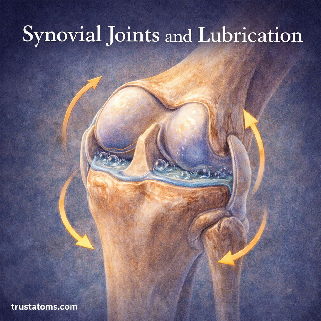
Synovial joints are the most common and allow the greatest range of motion. They have a fluid-filled joint cavity, articular cartilage covering the bone surfaces, and a surrounding joint capsule.
Examples include the shoulder, hip, knee, elbow, and wrist. Synovial fluid reduces friction and supports cartilage nutrition, allowing joint surfaces to move smoothly against one another.
Functional Classification: How Much Can a Joint Move?
Functional classification describes joints according to the degree of movement they permit rather than the material connecting their bones.
| Classification | Movement | Example |
|---|---|---|
| Synarthrosis | Little or no movement | Skull sutures |
| Amphiarthrosis | Slight movement | Pubic symphysis |
| Diarthrosis | Freely movable | Shoulder and knee |
Structural and functional classifications overlap but are not identical. For example, most fibrous skull sutures are synarthroses, while synovial joints are classified as diarthroses.
Six Types of Synovial Joints and Their Movements
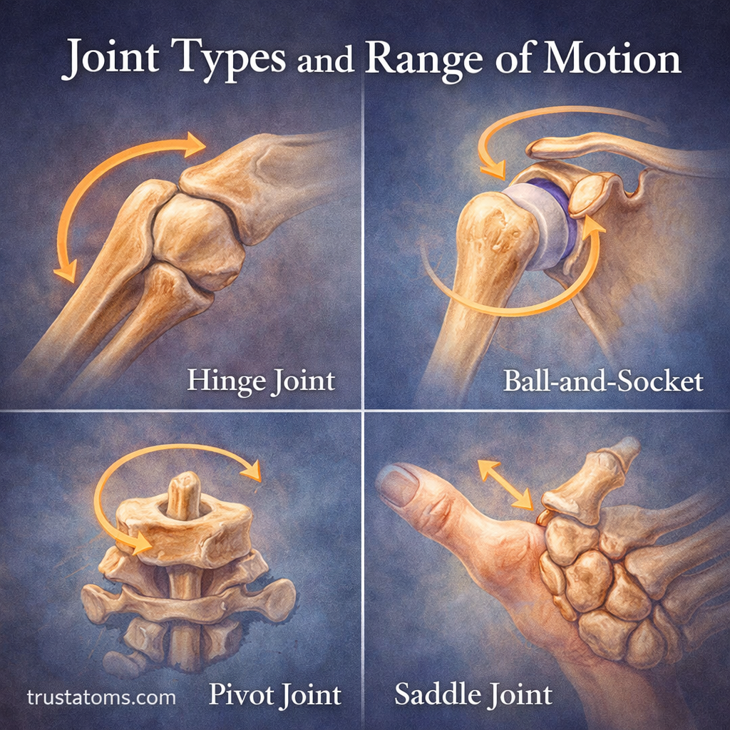
Synovial joints are divided into six types according to the shapes of their articulating surfaces and the directions in which they move.
1. Ball-and-Socket Joints
A rounded bone head fits into a socket, allowing movement around multiple axes. The shoulder and hip are examples.
These joints permit flexion, extension, abduction, adduction, internal and external rotation, and circumduction. The shoulder generally offers greater mobility, while the hip’s deeper socket provides more bony stability.
2. Hinge Joints
Hinge joints primarily permit flexion and extension, similar to the movement of a door hinge. The elbow is a clear example.
The knee is commonly classified as a modified hinge joint because it also permits a limited amount of rotation, particularly when flexed.
3. Pivot Joints
Pivot joints allow rotation around a single main axis. The atlantoaxial joint between the first two cervical vertebrae helps turn the head from side to side.
The proximal radioulnar joint is another example, allowing the radius to rotate during forearm pronation and supination.
4. Condyloid Joints
Also called ellipsoid joints, condyloid joints permit movement along two main axes. The radiocarpal joint of the wrist allows flexion, extension, radial deviation, and ulnar deviation.
These movements can combine into circumduction, but condyloid joints do not permit the same independent axial rotation as ball-and-socket joints.
5. Saddle Joints
Saddle joints have opposing surfaces that are concave in one direction and convex in another. The carpometacarpal joint at the base of the thumb is a familiar example.
This structure permits flexion, extension, abduction, adduction, and combined movements involved in thumb opposition, which allows the thumb to approach the fingertips.
6. Plane Joints
Plane joints, also called gliding joints, have relatively flat articular surfaces that permit small sliding movements. Examples include joints between certain wrist bones and between some bones of the foot.
Although movement at each individual plane joint is limited, the combined motion of several joints contributes to overall hand and foot flexibility.
What Is Range of Motion?
Range of motion (ROM) is the amount of movement available at a joint, usually measured in degrees. It depends on bone shape, soft-tissue flexibility, muscle activity, and joint condition.
- Active ROM: Movement produced by a person’s own muscle contractions.
- Passive ROM: Movement produced by an external force while the muscles being assessed remain relaxed.
- Functional ROM: The amount of movement needed to perform a particular everyday activity.
Example: Measuring Elbow Flexion
Imagine bending your elbow to bring a glass of water toward your mouth. An adult elbow commonly moves from approximately 0 degrees of extension to around 140–150 degrees of flexion, although normal values vary between individuals.
A clinician may use a goniometer, an instrument that measures joint angles, to assess this movement. If someone can actively bend their elbow to 120 degrees but reaches 140 degrees with gentle assistance, the difference may provide information about muscle performance, discomfort, or other movement limitations.
Common Joint Movement Terms
Anatomical movement terms describe the direction of motion rather than the specific joint involved.
- Flexion: Decreasing the angle between body segments, such as bending the elbow.
- Extension: Straightening a joint or increasing the angle between segments.
- Abduction: Moving a limb away from the body’s midline.
- Adduction: Moving a limb toward the body’s midline.
- Rotation: Turning a bone or body segment around its longitudinal axis.
- Circumduction: Combining movements so the distal end of a limb traces a roughly circular path.
Factors That Influence Joint Mobility
Joint movement varies naturally between people. Several factors determine the available range and how comfortably it can be used.
- Joint anatomy: Bone geometry determines which movements are mechanically possible.
- Muscles and connective tissues: Muscle length, tendon properties, and capsule stiffness can limit movement.
- Age and individual variation: Mobility differs with development, aging, genetics, and activity history.
- Injury or disease: Swelling, pain, arthritis, and scar tissue may restrict motion.
- Physical activity: Regular movement and appropriately performed flexibility exercises can help maintain or improve usable ROM.
More flexibility is not always better. Excessive joint laxity may reduce passive stability, while restricted motion can interfere with normal activities. The goal is sufficient mobility combined with effective muscular control.
How to Maintain Healthy Joint Movement
Joint health benefits from a combination of movement, strength, and appropriate recovery. Walking, resistance training, and controlled mobility exercises can help maintain functional movement and support surrounding tissues.
Gradually increasing activity is generally preferable to forcing a joint through painful or unfamiliar positions. Persistent pain, swelling, locking, or sudden loss of movement warrants evaluation by a qualified healthcare professional.
Frequently Asked Questions
Which joint type has the greatest range of motion?
Ball-and-socket joints allow movement around three axes and generally offer the greatest variety of movements. The shoulder has particularly extensive mobility because of its shallow socket and surrounding soft-tissue arrangement.
Why do some joints barely move?
Some joints are designed primarily for structural support and protection. Skull sutures, for example, form strong fibrous connections that help protect the brain rather than allowing significant movement between cranial bones.
Can range of motion improve with exercise?
Yes. Regular stretching, strengthening, and controlled movement can improve or maintain range of motion in many joints. However, bone structure and certain medical conditions impose limits that exercise cannot eliminate.
Joint types explain why the skull remains stable, the elbow bends mainly in one plane, and the shoulder moves in several directions. Structural design establishes each joint’s movement possibilities, while muscles and connective tissues influence how that movement is controlled. Understanding these differences provides a practical foundation for studying mobility, flexibility, and musculoskeletal function.
This article is for general education and is not medical advice. Talk to a qualified health professional about any medical concern.
Sources: OpenStax, Anatomy and Physiology 2e; Gray’s Anatomy for Students; American Academy of Orthopaedic Surgeons, OrthoInfo.