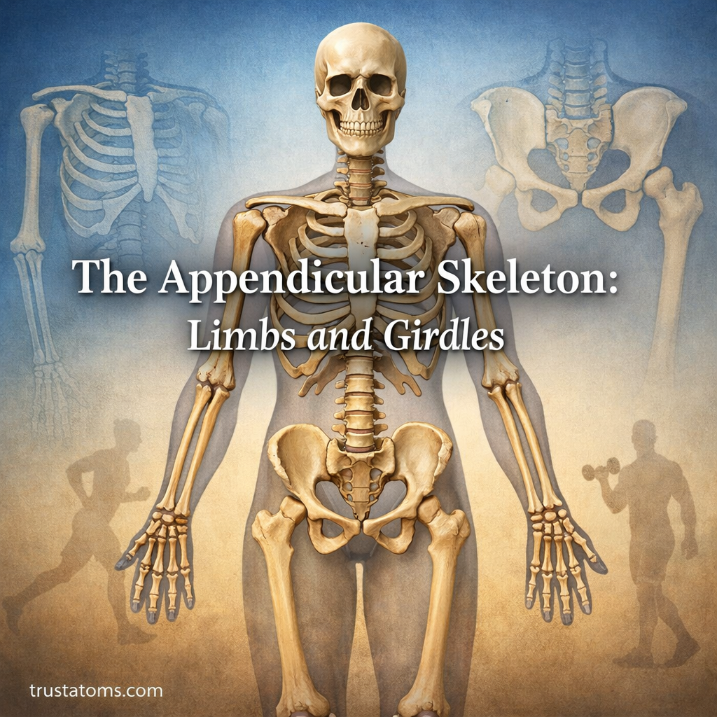
The appendicular skeleton consists of 126 bones that form the arms, hands, legs, feet, shoulder girdles, and pelvic girdle of the typical adult human skeleton. These bones support movement, provide attachment points for muscles, and connect the limbs to the body’s central skeletal framework.
In human anatomy, the skeleton is divided into axial and appendicular regions. While the axial skeleton forms the body’s central structure, the appendicular skeleton includes the limbs and the girdles that attach them to the core, allowing walking, lifting, grasping, and other movements.
What Is the Appendicular Skeleton?
The appendicular skeleton includes four main regions: the pectoral girdles, upper limbs, pelvic girdle, and lower limbs. Together, these structures account for 126 of the 206 bones typically found in the adult skeleton.
| Region | Number of Bones | Main Function |
|---|---|---|
| Pectoral girdles | 4 | Attach and position the upper limbs |
| Upper limbs | 60 | Reaching, grasping, and manipulation |
| Pelvic girdle | 2 | Transfer weight and support the hips |
| Lower limbs | 60 | Weight-bearing and locomotion |
| Total | 126 | Movement and skeletal support |
These bones operate with muscles, tendons, ligaments, and joints as part of the Musculoskeletal System. Their functions can be seen in Hip Joint Mechanics and Stability and Knee Joint Mechanics, where forces must be transferred while maintaining controlled movement.
The Pectoral Girdle: Connecting the Arms to the Trunk
Each pectoral girdle consists of two bones: a clavicle (collarbone) and a scapula (shoulder blade). Together, the left and right pectoral girdles contain four bones.
The clavicle connects the upper limb to the axial skeleton through the sternoclavicular joint. The scapula provides attachment sites for numerous muscles and contains the glenoid cavity, which articulates with the head of the humerus to form the shoulder joint.
Unlike the pelvic girdle, the shoulder region relies heavily on muscles for stability. This arrangement allows extensive movement but also makes the shoulder vulnerable to instability and dislocation. The relationship between flexibility and stability is explored in Shoulder Joint Mechanics and Mobility.
The Upper Limbs: Arms, Wrists, and Hands
Each upper limb contains 30 bones, giving both arms and hands a combined total of 60 bones. These structures allow powerful movements as well as precise manipulation of objects.
- Humerus: The single bone of the upper arm, connecting the shoulder to the elbow.
- Radius: The forearm bone on the thumb side, involved in wrist movement and forearm rotation.
- Ulna: The forearm bone on the little-finger side, forming a major part of the elbow joint.
- Carpals: Eight small bones forming the wrist.
- Metacarpals: Five bones forming the framework of the palm.
- Phalanges: Fourteen finger bones, including two in the thumb and three in each other finger.
The radius rotates around the ulna during pronation and supination, allowing the palm to turn downward or upward. Combined with wrist and finger movements, this makes activities such as writing, typing, and using tools possible.
The Pelvic Girdle: Supporting Body Weight
The pelvic girdle consists of two hip bones, also called coxal bones. Each hip bone develops from three regions that fuse during maturation: the ilium, ischium, and pubis.
The hip bones articulate with the sacrum at the sacroiliac joints and meet anteriorly at the pubic symphysis. Together with the sacrum and coccyx, they form the bony pelvis, although the sacrum and coccyx belong to the axial skeleton.
Each hip bone contains a deep socket called the acetabulum, which receives the head of the femur. This arrangement creates a stable ball-and-socket joint capable of supporting substantial loads during standing, walking, and running.
Compared with the shoulder girdle, the pelvic girdle has stronger skeletal connections to the trunk. Its structure helps transfer forces between the lower limbs and spine while supporting pelvic organs.
The Lower Limbs: Legs, Ankles, and Feet
Each lower limb contains 30 bones, including those of the thigh, knee, leg, ankle, and foot. The lower limbs are adapted to support body weight, absorb forces, and generate movement during locomotion.
- Femur: The thigh bone and the longest bone in the human body.
- Patella: The kneecap, which improves the mechanical advantage of the quadriceps muscle.
- Tibia: The larger, medial leg bone that bears most of the load transmitted through the lower leg.
- Fibula: The slender, lateral leg bone that provides muscle attachments and contributes to ankle stability.
- Tarsals: Seven bones forming the ankle and rear portion of the foot.
- Metatarsals: Five bones connecting the tarsals to the toes.
- Phalanges: Fourteen toe bones, including two in the big toe.
The arches of the feet help distribute forces and provide both flexibility and support. The ankle, knee, and hip work together to maintain balance and move the body efficiently.
How the Appendicular Skeleton Produces Movement
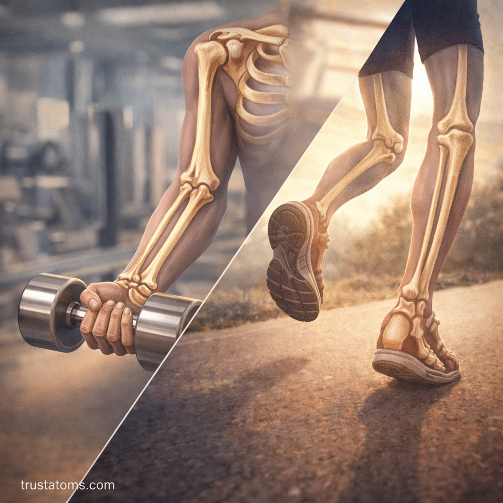
Movement occurs when skeletal muscles contract and pull on bones through tendons. Joints permit controlled movement, while bones function as levers that transmit forces and produce changes in position.
For example, when lifting a dumbbell, the biceps brachii helps bend the elbow by pulling on the radius through its tendon. During running, muscles around the hips, knees, and ankles coordinate to support body weight and propel the body forward.
Several joint types make these movements possible:
- Ball-and-socket joints: The shoulder and hip allow movement in multiple directions, including rotation.
- Hinge joints: The elbow and knee primarily permit bending and straightening, although the knee also allows limited rotation.
- Pivot joints: The proximal and distal radioulnar joints allow rotation of the forearm.
- Condyloid joints: The wrist and metacarpophalangeal joints permit movement along two principal axes.
Upper vs. Lower Limbs: Functional Differences
The upper and lower limbs share a similar general arrangement of bones, but their structures reflect different mechanical demands.
| Feature | Upper Limbs | Lower Limbs |
|---|---|---|
| Primary function | Manipulation and reaching | Weight support and locomotion |
| Girdle | Pectoral girdle | Pelvic girdle |
| Major proximal joint | Shoulder | Hip |
| Typical mechanical priority | Mobility and precision | Stability and force transmission |
| Distal specialization | Grasping with hands | Balance and propulsion with feet |
Common Injuries and Skeletal Conditions
Because appendicular bones and joints experience repeated mechanical loading, they can be affected by fractures, dislocations, ligament injuries, and degenerative conditions.
- Fractures: Breaks in bones caused by trauma, repetitive stress, or weakened bone tissue.
- Dislocations: Displacement of bones at a joint, commonly affecting the shoulder.
- Sprains: Stretching or tearing of ligaments that stabilize joints.
- Osteoarthritis: Degeneration affecting joint cartilage and other joint tissues, often involving the knees, hips, or hands.
Maintaining muscle strength, balance, and appropriate physical activity supports the appendicular skeleton. Adequate calcium and vitamin D are also necessary for normal bone maintenance.
Frequently Asked Questions
How many bones are in the appendicular skeleton?
The typical adult appendicular skeleton contains 126 bones: 4 in the pectoral girdles, 60 in the upper limbs, 2 in the pelvic girdle, and 60 in the lower limbs. Individual bone counts can vary because of anatomical variations.
Is the pelvis part of the appendicular skeleton?
The two hip bones belong to the appendicular skeleton and form the pelvic girdle. The sacrum and coccyx also contribute to the bony pelvis but are classified as part of the axial skeleton.
What is the difference between the axial and appendicular skeleton?
The axial skeleton forms the central framework of the body, including the skull, vertebral column, ribs, and sternum. The appendicular skeleton consists of the limbs and their girdles, supporting movement and interaction with the environment.
The appendicular skeleton combines 126 bones into a framework specialized for movement, weight-bearing, and manipulation. Its shoulder and pelvic girdles connect the limbs to the axial skeleton, while joints and muscles allow everything from precise finger movements to walking and running.
This article is for general education and is not medical advice. Talk to a qualified health professional about any medical concern.
Sources: Gray’s Anatomy: The Anatomical Basis of Clinical Practice; OpenStax Anatomy and Physiology 2e; Moore’s Clinically Oriented Anatomy.