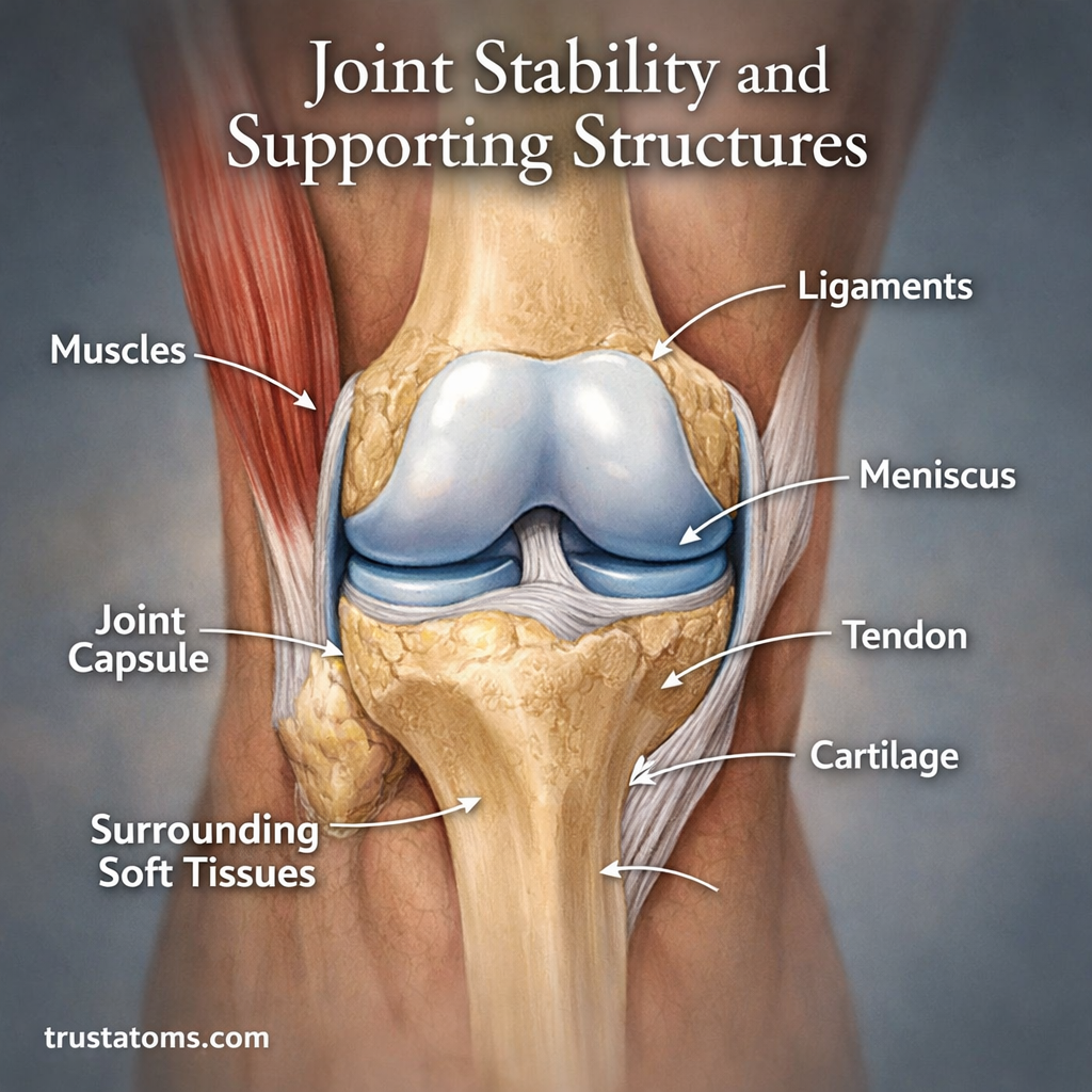
Joint stability is the ability of a joint to maintain controlled alignment and resist excessive movement under physical forces. Bones, ligaments, muscles, tendons, joint capsules, and sensory feedback work together to keep joints supported while allowing normal movement.
Understanding joint stability is an important part of human anatomy because every activity, from walking downstairs to lifting a grocery bag, requires a balance between mobility and support. Different joints achieve that balance through different combinations of structural and muscular control.
What Is Joint Stability?
Joint stability describes how effectively a joint maintains appropriate contact and alignment between its bones while responding to movement and external loads. A stable joint can move through its normal range without unwanted shifting, excessive separation, or loss of control.
Stability does not mean stiffness. Healthy joints must permit movement while resisting forces that could damage supporting tissues. The amount of stability required depends on the joint’s shape, function, and typical loading conditions.
For example, Hip Joint Mechanics and Stability explains how a deep socket supports weight-bearing movement. In contrast, Shoulder Joint Mechanics and Mobility describes a joint that sacrifices some bony stability to achieve a wider range of motion.
How Joint Shape Affects Stability and Mobility
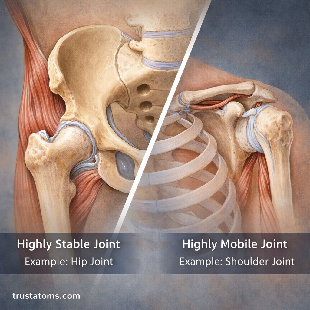
Joint geometry influences how securely bones fit together. A deep socket provides more bony containment, while a shallow socket permits greater freedom of movement but generally requires more soft-tissue support.
- Hip: The femoral head fits into the deep acetabulum, providing substantial stability during standing, walking, and weight-bearing.
- Shoulder: The relatively shallow glenoid socket allows extensive arm movement, with muscles, ligaments, and the labrum helping maintain stability.
- Knee: The joint permits mainly bending and straightening, with controlled rotation supported by ligaments, menisci, and surrounding muscles.
Most freely movable joints are synovial joints. Their capsules, cartilage-covered surfaces, and lubricating fluid are explained further in Synovial Joints and Lubrication.
Six Structures That Support Joint Stability
Ligaments Limit Excessive Movement
Ligaments are strong bands of connective tissue connecting bone to bone. They guide joint movement and resist excessive stretching, twisting, or separation.
At the knee, the anterior cruciate ligament helps resist forward movement of the tibia relative to the femur. Other ligaments resist different forces, allowing the joint to remain controlled during changing movements.
Tendons Transfer Muscle Forces
Tendons connect muscles to bones and contribute to joint stability through movement. When muscles contract, tendons transmit their forces to the skeleton, helping position and control the joint.
Unlike ligaments, which primarily provide passive restraint, tendons support stability through the actions of the muscles attached to them.
Muscles Provide Active Control
Muscles adjust their force as the body changes position. They can stabilize a joint before movement begins, control motion while it occurs, and respond to unexpected disturbances.
The rotator cuff muscles, for example, help keep the upper arm bone centered against the shoulder socket during reaching and lifting.
Joint Capsules Reinforce the Joint
A synovial joint capsule surrounds the joint cavity. Its outer fibrous layer helps hold the bones together and restrict excessive motion, while its inner synovial membrane produces synovial fluid.
Cartilage Distributes Contact Forces
Cartilage covers the ends of bones within joints. Articular cartilage creates a smooth, low-friction surface and helps distribute contact forces during movement.
Additional fibrocartilage structures, such as the knee menisci and shoulder labrum, can improve joint congruence and load distribution. Cartilage supports stability indirectly by helping joint surfaces interact effectively.
Synovial Fluid Reduces Friction
Synovial fluid lubricates joint surfaces and helps nourish articular cartilage. Although it does not act as a primary mechanical restraint, it supports smooth movement and normal joint function.
Static Stability Versus Dynamic Stability
Joint stability is commonly described in terms of passive structural support and active muscular control. These systems operate together rather than independently.
| Feature | Static Stability | Dynamic Stability |
|---|---|---|
| Main contributors | Bones, ligaments, capsules, and other passive tissues | Muscles and their tendons |
| How it works | Resists movement through structural shape and tissue tension | Adjusts muscular forces to control movement |
| Example | Knee ligaments limiting excessive translation | Quadriceps and hamstrings controlling knee position |
| Response | Depends on joint position and applied forces | Changes with muscle activation and coordination |
Neither system can fully replace the other. Strong muscles may compensate for some limitations in passive structures, but severe ligament damage can still produce mechanical instability.
How the Nervous System Helps Stabilize Joints
Joint control also depends on proprioception, the body’s ability to sense position and movement. Sensory receptors in muscles, tendons, joint tissues, and skin send information to the nervous system.
The nervous system uses this feedback, together with planned movement commands, to coordinate muscle activity and maintain balance.
Example: Stepping Onto Uneven Ground
Imagine walking onto a slightly tilted sidewalk. As your foot contacts the uneven surface, sensory information helps your nervous system detect the changing position of the ankle.
Muscles around the ankle and lower leg adjust their activity to control the joint and maintain balance. Ligaments provide additional passive resistance if the ankle approaches the limits of its normal motion. This combination of sensory feedback, muscle control, and structural restraint helps prevent unwanted movement, although it cannot prevent every sprain.
Factors That Influence Joint Stability
Stability varies between individuals and can change with physical condition, activity, and injury history.
- Bone shape: Joint surface depth and geometry influence how well bones remain aligned.
- Ligament integrity: Injured or unusually lax ligaments may permit excessive movement.
- Muscle capacity: Strength, endurance, and timely activation affect dynamic control.
- Joint mobility: Adequate range of motion supports function, while excessive laxity can contribute to instability in some individuals.
- Fatigue and coordination: Tired muscles or impaired sensory control can reduce movement precision.
- Previous injuries: Ligament injuries and dislocations can alter mechanical restraint or proprioception.
Common Problems Caused by Joint Instability
When supporting structures cannot adequately control movement, joints may become more vulnerable to injury or repeated episodes of giving way.
- Sprains: Ligaments become stretched or torn when forces exceed their tolerance.
- Dislocations: Joint surfaces lose their normal contact, often damaging surrounding tissues.
- Chronic instability: Recurrent giving way may follow ligament injury, repeated dislocations, or impaired muscular control.
- Secondary tissue irritation: Altered movement patterns may increase stress on cartilage, tendons, or other structures.
Joint pain and instability are not identical. A painful joint may remain mechanically stable, while some unstable joints produce relatively little pain between episodes.
Ways to Improve and Maintain Joint Stability
Exercise can improve the muscular and neurological components of joint stability. The most useful approach depends on the joint involved, current physical capacity, and any existing injury.
- Progressive strength training: Build the capacity of muscles surrounding the joints through manageable resistance exercises.
- Balance exercises: Practice controlled single-leg standing or similar activities appropriate to your ability.
- Movement coordination: Develop control during stepping, turning, reaching, and changes of direction.
- Appropriate mobility work: Maintain functional movement without forcing joints beyond comfortable limits.
- Gradual activity progression: Increase training demands progressively and allow sufficient recovery.
After significant ligament injuries or repeated dislocations, rehabilitation may require individualized strengthening, balance training, and medical assessment. Exercise improves active support but does not necessarily restore a torn ligament or correct every structural cause of instability.
Frequently Asked Questions
What is the difference between joint stability and mobility?
Stability is the ability to maintain controlled alignment and resist unwanted movement. Mobility is the ability to move through an available range of motion. Healthy joint function requires an appropriate combination of both rather than maximizing either one.
Can stronger muscles compensate for weak ligaments?
Stronger, well-coordinated muscles can improve dynamic stability and sometimes compensate for reduced ligament support. However, muscles cannot completely replace the passive restraint of severely damaged ligaments, particularly during sudden or high-force movements.
Are flexible joints always less stable?
No. A large range of motion does not automatically mean a joint is unstable. Some people have naturally flexible joints with excellent muscular control. Instability becomes a concern when excessive movement causes symptoms, repeated giving way, or difficulty controlling joint position.
Joint stability results from the combined effects of bone geometry, connective tissues, muscular forces, and nervous system coordination. The hip, shoulder, and knee demonstrate how different anatomical designs balance movement with mechanical restraint. Maintaining strength, coordination, and appropriate mobility helps these supporting systems work together during everyday activities.
This article is for general education and is not medical advice. Talk to a qualified health professional about any medical concern.
Sources: Gray’s Anatomy: The Anatomical Basis of Clinical Practice; OpenStax, Anatomy and Physiology 2e; American Academy of Orthopaedic Surgeons, OrthoInfo.