Digestive sphincters are specialized muscular structures that regulate when food, digestive fluids, and waste move between different parts of the gastrointestinal tract. By contracting and relaxing at specific times, they coordinate swallowing, stomach emptying, intestinal flow, and bowel movements while helping prevent unwanted backflow.
These structures are an example of how human anatomy supports coordinated organ function. When you swallow a bite of food, several muscles must open and close in sequence so the food reaches your stomach without moving backward into your throat.
What Are Digestive Sphincters?
A sphincter is a ring-like muscular structure that controls an opening or passage. Many digestive sphincters contain smooth muscle, which operates involuntarily, while others contain skeletal muscle that allows more direct voluntary control.
Some sphincters are clearly defined anatomical rings. Others, such as the lower esophageal sphincter, are functional high-pressure zones maintained by muscle activity and surrounding structures.
Their main functions include regulating forward movement, limiting reflux, protecting sensitive tissues, and controlling the timing of digestive processes.
Sphincter activity is especially noticeable during swallowing, when coordinated relaxation permits food to enter the esophagus and stomach. The sequence is explained further in swallowing mechanics and esophageal transport.
Major Sphincters in the Digestive System
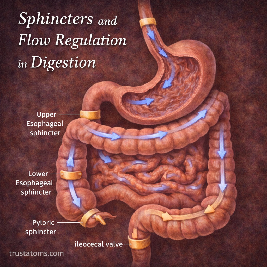
Several important control points are distributed along the digestive tract. Their locations can be understood through digestive tract anatomy, which follows food from the mouth to the colon.
Sphincters work alongside muscular contractions that propel digestive contents forward. Gut motility and peristalsis explains how these contractions move material between control points.
Upper Esophageal Sphincter
The upper esophageal sphincter (UES) lies between the pharynx and esophagus. It normally remains closed, helping prevent air from entering the esophagus during breathing and limiting the backward movement of esophageal contents toward the throat.
During swallowing, it briefly relaxes while surrounding muscles help open the passage. It then closes again after the swallowed material enters the esophagus.
Lower Esophageal Sphincter
Located between the esophagus and stomach, the lower esophageal sphincter (LES) normally maintains a pressure barrier that helps prevent acidic stomach contents from flowing upward.
It relaxes during swallowing to let food enter the stomach. The diaphragm also contributes to the anti-reflux barrier around this junction. If the barrier becomes ineffective or relaxes inappropriately, stomach contents may reflux into the esophagus.
Pyloric Sphincter
The pyloric sphincter sits between the stomach and the duodenum, the first section of the small intestine. It helps regulate the passage of chyme, the mixture of partially digested food and digestive fluids.
Stomach contractions push chyme toward the pylorus, but much of the material is propelled backward for further mixing. Small amounts pass into the duodenum as the pyloric region coordinates with stomach and intestinal activity.
Ileocecal Valve
The ileocecal valve is located where the ileum, the final section of the small intestine, joins the cecum of the large intestine.
Its structure and associated muscular activity help regulate the movement of intestinal contents into the colon and limit backward flow from the bacteria-rich large intestine into the small intestine.
Internal and External Anal Sphincters
Two sphincters contribute to bowel control at the end of the digestive tract:
- Internal anal sphincter: Made of smooth muscle and controlled involuntarily, it maintains resting tension that helps prevent leakage.
- External anal sphincter: Made of skeletal muscle and subject to voluntary control, it helps delay defecation until an appropriate time.
The puborectalis muscle and pelvic floor also contribute to continence by supporting the anorectal angle and coordinating with the anal sphincters.
How Sphincters Coordinate Food Movement
Digestive sphincters do not simply open one after another. Their activity is coordinated with muscular contractions, pressure changes, and signals from nearby organs.
- Swallowing: The upper esophageal sphincter opens to admit food into the esophagus.
- Esophageal transport: Peristaltic contractions move food toward the stomach as the lower esophageal sphincter relaxes.
- Stomach emptying: The pyloric region regulates the gradual delivery of chyme into the duodenum.
- Intestinal passage: The ileocecal valve helps control the transfer of contents into the colon.
- Defecation: The anal sphincters and pelvic floor coordinate to permit or delay the release of stool.
This coordination allows different digestive regions to perform their functions without receiving more material than they can process efficiently.
What Controls Sphincter Function?
Digestive sphincters respond to signals from the nervous system, local digestive reflexes, chemical messengers, and changes in pressure.
Enteric and Autonomic Nerves
The enteric nervous system, a network of neurons within the gastrointestinal wall, helps coordinate muscle contractions and relaxation. Parasympathetic and sympathetic pathways modify this activity according to digestive conditions.
For example, inhibitory nerve signals help the lower esophageal sphincter relax during swallowing. Other neural circuits regulate the pylorus and coordinate reflexes involved in bowel movements.
Hormones and Local Reflexes
Digestive hormones and local signaling molecules influence the movement of food through the stomach and intestines. Hormones such as cholecystokinin and secretin can help slow gastric emptying when nutrients or acid enter the duodenum.
Stretch receptors also detect changes in pressure and fullness. Distension of the rectum, for instance, triggers a reflex that relaxes the internal anal sphincter while other muscles help maintain continence until defecation is appropriate.
As material reaches the colon, these control mechanisms interact with the processes described in large intestine function and water absorption.
What Happens When Digestive Sphincters Malfunction?
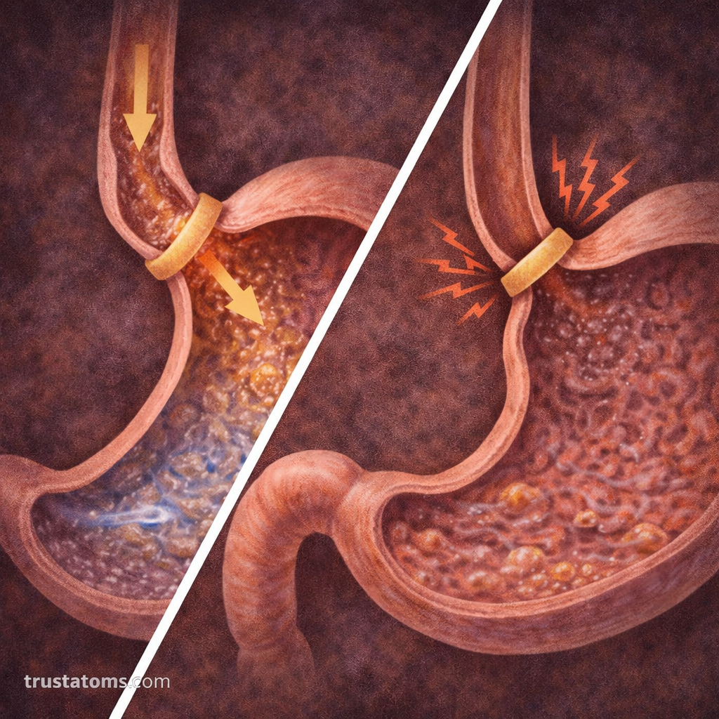
When a sphincter fails to relax, contracts abnormally, or cannot maintain sufficient closure, digestive flow may be disrupted.
- Gastroesophageal reflux: An ineffective anti-reflux barrier allows stomach contents to enter the esophagus, potentially causing heartburn or regurgitation.
- Achalasia: Impaired relaxation of the lower esophageal sphincter, combined with abnormal esophageal motility, makes swallowing difficult.
- Pyloric stenosis: Narrowing at the pylorus restricts gastric emptying and may cause vomiting.
- Fecal incontinence: Weakness or impaired coordination of anal sphincters, nerves, and pelvic floor muscles can contribute to loss of bowel control.
Not every digestive symptom indicates a sphincter disorder. Delayed stomach emptying, for example, can arise from several causes unrelated to physical narrowing of the pylorus. Persistent difficulty swallowing, recurrent vomiting, or loss of bowel control should be evaluated by a qualified healthcare professional.
How Sphincters Differ From Valves
A sphincter controls an opening primarily through muscular contraction and relaxation. A valve may regulate flow through its shape, tissue folds, pressure differences, or associated muscular activity.
The ileocecal valve illustrates this overlap because its structural features and muscular components work together. In contrast, the lower esophageal sphincter is best understood as a functional pressure barrier rather than a simple flap-like valve.
Frequently Asked Questions
Are digestive sphincters controlled voluntarily?
Most digestive sphincters operate involuntarily through smooth muscle and neural reflexes. The external anal sphincter contains skeletal muscle and can be voluntarily contracted to help delay a bowel movement.
Which sphincter prevents stomach acid from entering the esophagus?
The lower esophageal sphincter helps prevent acid reflux by maintaining a pressure barrier between the stomach and esophagus. The surrounding diaphragm and anatomy of the gastroesophageal junction also contribute to this protection.
What controls how quickly food leaves the stomach?
Gastric emptying depends on coordinated contractions of the stomach, pyloric activity, and feedback from the duodenum. The composition and physical characteristics of a meal also influence how quickly its contents enter the small intestine.
Digestive sphincters coordinate movement between the esophagus, stomach, intestines, and rectum by controlling when passages open and close. Their activity works with peristalsis, local reflexes, and nervous system signals to regulate flow and reduce unwanted backflow. When these mechanisms become disrupted, the effects can range from reflux and swallowing difficulties to impaired stomach emptying or bowel control.
This article is for general education and is not medical advice. Talk to a qualified health professional about any medical concern.
Sources: OpenStax, Anatomy and Physiology 2e; Hall and Hall, Guyton and Hall Textbook of Medical Physiology; National Institute of Diabetes and Digestive and Kidney Diseases.