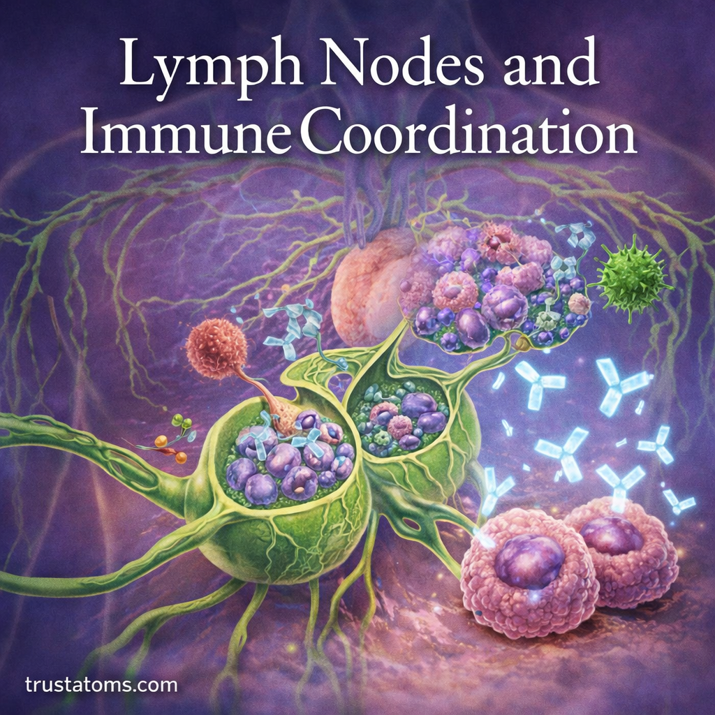
Lymph nodes are small, bean-shaped organs that filter lymph fluid and coordinate immune responses against infections and other threats. They bring immune cells together, allowing the body to recognize foreign substances, activate targeted defenses, and develop immune memory.
Found throughout the body, lymph nodes connect the movement of tissue fluid with the activity of the immune system. Understanding their structure and function is an essential part of studying human anatomy and how the body protects itself from disease.
What Are Lymph Nodes?
Lymph nodes are part of the lymphatic system—a network of vessels, tissues, and organs that transport lymph fluid throughout the body. Lymph forms when excess fluid from surrounding tissues enters lymphatic vessels.
Hundreds of lymph nodes are distributed throughout the body, often grouped near major lymphatic pathways. Common locations include the neck, armpits, groin, chest, and abdomen.
Each lymph node contains immune cells that monitor lymph fluid for pathogens like bacteria, viruses, and abnormal cells. These cells recognize suspicious materials and help initiate appropriate immune responses.
For a closer look at how lymph travels between tissues and lymph nodes, see Lymph Vessels and Lymph Nodes Overview.
Structure of a Lymph Node
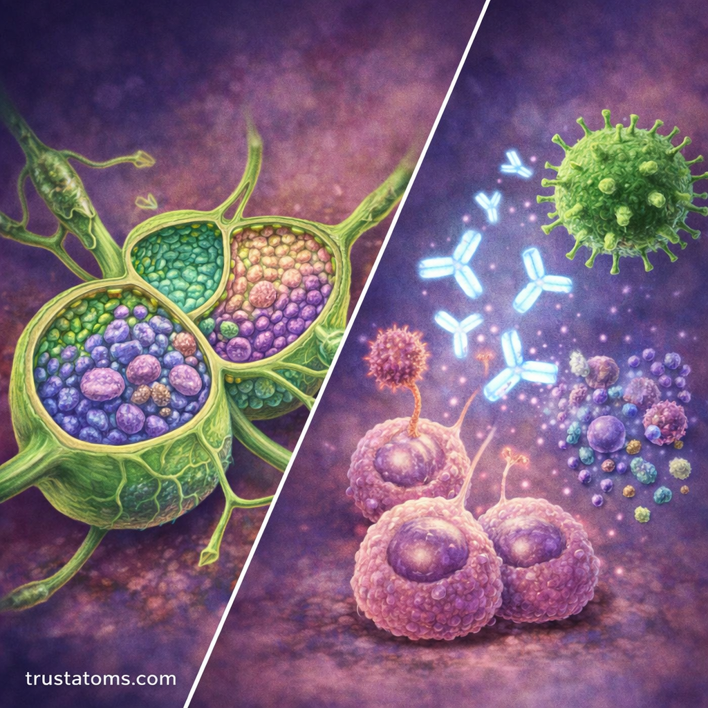
A lymph node is surrounded by a connective tissue capsule and divided into specialized regions. This arrangement helps immune cells encounter antigens, communicate, and respond efficiently.
| Region | Main Features | Primary Function |
|---|---|---|
| Cortex | B-cell follicles, including germinal centers in activated follicles | Supports B-cell activation and antibody-related responses |
| Paracortex | T cells and dendritic cells | Supports antigen presentation and T-cell activation |
| Medulla | Medullary cords and sinuses containing plasma cells and macrophages | Supports antibody secretion and filtration |
Lymph enters through vessels on the outer surface and moves through internal channels called sinuses. It eventually leaves through an efferent lymphatic vessel at the hilum, where blood vessels also enter and exit.
How Lymph Nodes Filter Lymph Fluid
As lymph moves through a node, its flow slows, allowing immune cells to examine materials carried from surrounding tissues. The filtering process follows several stages:
- Entry: Lymph arrives through afferent lymphatic vessels.
- Movement: Fluid passes through subcapsular, cortical, and medullary sinuses.
- Detection: Macrophages and other immune cells encounter foreign particles and antigens.
- Response: Some materials are engulfed, while antigens may trigger lymphocyte activation.
- Exit: Filtered lymph leaves through an efferent lymphatic vessel.
This process helps remove microorganisms and cellular debris, although filtration does not guarantee that every pathogen or abnormal cell will be eliminated.
Role of Lymph Nodes in Immune Coordination
Lymph nodes function as meeting places for antigen-presenting cells and lymphocytes. They connect early detection of potential threats with the more specialized responses of adaptive immunity.
Several types of white blood cells participate in this coordination. Their broader defensive functions are explained in White Blood Cells and Immune Defense.
How T Cells and B Cells Become Activated
Dendritic cells can collect antigens in infected tissues and carry them to nearby lymph nodes. There, they display antigen fragments to T cells, potentially activating T cells that recognize the presented material and receive the necessary additional signals.
B cells recognize specific antigens through receptors on their surfaces. With appropriate signals, often including assistance from helper T cells, activated B cells can multiply and develop into antibody-secreting plasma cells or memory B cells.
Some activated T cells also become long-lived memory cells. These populations help the immune system respond more effectively when it encounters the same pathogen again.
How Lymph Nodes Differ From the Spleen
Lymph nodes primarily monitor lymph draining from tissues, while the spleen monitors blood and helps remove aging blood cells and certain bloodborne microorganisms. This distinction is explored further in Spleen Function in Immune Filtering.
Why Lymph Nodes Swell During Infection
Enlarged lymph nodes, known as lymphadenopathy, often occur when immune activity increases. Activated lymphocytes multiply, additional immune cells enter the node, and inflammation can cause the tissue to expand.
For example, during a throat infection, lymph nodes along the sides of the neck may become swollen and tender. These nodes receive lymph from nearby tissues and may respond to antigens associated with the infection.
Swelling can also result from inflammatory disorders, medications, and certain cancers. Enlarged nodes are not automatically evidence of a serious disease, but persistent, unexplained, enlarging, or unusually hard nodes warrant medical evaluation, particularly when accompanied by fever, night sweats, or unexplained weight loss.
How Doctors Examine Lymph Nodes
Healthcare professionals may examine lymph nodes to investigate infections, inflammatory conditions, or possible cancer spread. Evaluation begins with medical history and a physical examination, including the nodes’ location, size, tenderness, and mobility.
When additional investigation is needed, clinicians may use ultrasound or other imaging techniques. A needle sample or surgical biopsy can provide cells or tissue for laboratory examination.
In cancer care, nearby lymph nodes may be examined to determine whether malignant cells have spread from a primary tumor. Sentinel lymph node biopsy, used for selected cancers, examines the first node or nodes most likely to receive drainage from the tumor region.
Lymph Nodes and Other Immune Organs
Lymph nodes work alongside several other organs involved in immune cell development and defense:
- Bone marrow: Produces blood cells and supports B-cell development.
- Thymus: Supports T-cell maturation and selection.
- Spleen: Filters blood and supports responses to bloodborne antigens.
- Tonsils and other mucosal lymphoid tissues: Help monitor materials entering through mucosal surfaces.
Together, these structures provide different locations for immune cell development, antigen detection, and coordinated defense.
Frequently Asked Questions
Where are most noticeable lymph nodes located?
Clusters near the neck, armpits, and groin are among the easiest to examine because they lie relatively close to the skin. Many other lymph nodes are located deeper in the chest and abdomen and cannot normally be felt.
Does a swollen lymph node always mean an infection?
No. Infection is a common cause, but immune disorders, inflammatory conditions, medications, and cancers can also cause enlargement. The significance depends on the node’s characteristics, accompanying symptoms, and medical history.
Do lymph nodes produce antibodies?
Antibodies are produced by plasma cells, some of which develop from activated B cells within lymph nodes. The nodes provide an organized environment where B cells receive signals, multiply, and differentiate into antibody-secreting cells.
Lymph nodes connect tissue drainage with immune surveillance by filtering lymph and organizing encounters between antigens and immune cells. Their specialized internal regions support T-cell activation, B-cell responses, and immune memory. Understanding these processes also explains why lymph nodes can enlarge during infection and why clinicians examine them when investigating disease.
This article is for general education and is not medical advice. Talk to a qualified health professional about any medical concern.
Sources: Abbas, Lichtman, and Pillai, Cellular and Molecular Immunology; Ross and Pawlina, Histology: A Text and Atlas; National Cancer Institute.