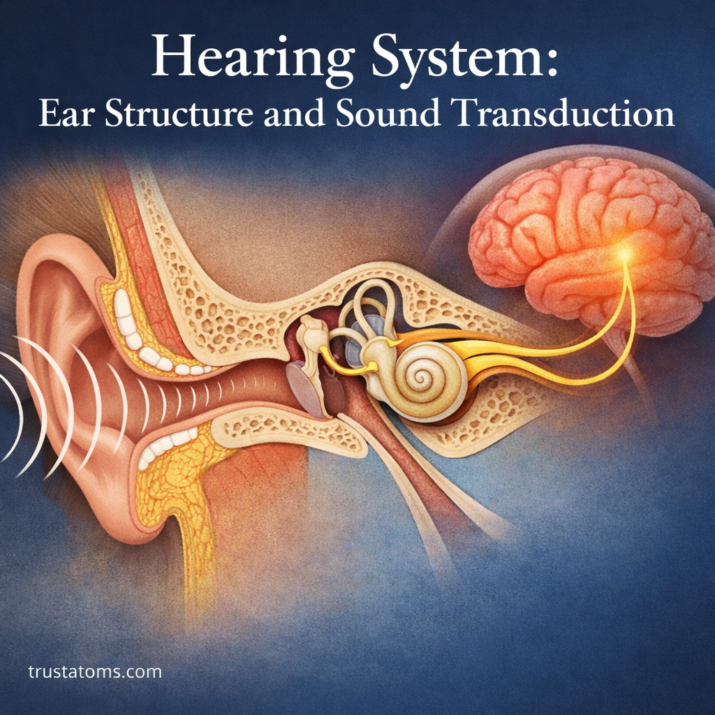
The human hearing system detects sound waves and converts their mechanical vibrations into electrical signals that the brain interprets as sound. This process, called sound transduction, involves the outer ear collecting sound, the middle ear transmitting vibrations, and sensory hair cells in the inner ear activating auditory nerve fibers.
Hearing demonstrates how structures studied in human anatomy work with the nervous system to produce sensory experiences. From recognizing speech to locating an approaching vehicle, hearing depends on accurate communication between the ears and brain.
How the Hearing System Detects and Processes Sound
Hearing involves three connected components: the ear, the auditory nerve, and specialized processing centers in the brain. Sound begins as pressure variations traveling through air and becomes meaningful only after the nervous system interprets the resulting signals.
The hearing system operates through several stages:
- Sound collection: The outer ear directs incoming sound toward the eardrum.
- Mechanical transmission: The middle ear transfers vibrations into the inner ear.
- Sensory transduction: Cochlear hair cells convert movement into electrical responses.
- Neural processing: Auditory pathways transmit information that the brain uses to recognize sound.
Hearing and vision both depend on specialized sensory receptors, although they detect different forms of energy. The vision system and light processing pathway converts light into neural signals rather than mechanical vibrations.
The Three Main Sections of the Ear
The ear is divided into outer, middle, and inner regions. Each has a distinct function in transmitting or detecting sound.
Outer Ear: Collecting Sound Waves
The outer ear includes the pinna, also called the auricle, and the external auditory canal. The pinna helps collect sound and provides directional cues, while the ear canal carries sound toward the tympanic membrane, commonly called the eardrum.
Middle Ear: Transmitting Vibrations
The middle ear is an air-filled cavity containing three small bones called the auditory ossicles:
- Malleus: The hammer-shaped bone attached to the eardrum.
- Incus: The anvil-shaped bone connecting the malleus and stapes.
- Stapes: The stirrup-shaped bone that transfers vibrations to the oval window of the inner ear.
The ossicles increase pressure at the oval window, helping transfer sound energy from air into cochlear fluid. The Eustachian tube connects the middle ear to the upper throat and helps equalize air pressure across the eardrum.
Inner Ear: Detecting Sound and Movement
The inner ear contains the cochlea, a spiral-shaped, fluid-filled structure responsible for hearing. Inside it, the organ of Corti contains sensory hair cells that respond to mechanical movement.
The semicircular canals are primarily involved in balance and the vestibular system, not hearing, but they are part of the inner ear. Together with the otolith organs, they detect head movement and orientation.
The Path Sound Takes Through the Ear
Sound travels through a sequence of structures before activating the auditory nerve.
- Sound enters the ear canal: Air pressure variations reach the eardrum.
- The eardrum vibrates: Its movement follows changes in the incoming sound wave.
- The ossicles transmit motion: The malleus, incus, and stapes transfer vibrations toward the inner ear.
- The oval window moves: Stapes movement creates pressure waves in cochlear fluid.
- The basilar membrane vibrates: Different regions respond most strongly to different sound frequencies.
- Hair cells respond: Mechanical deflection of their sensory projections produces electrical changes that lead to auditory nerve activity.
This sequence allows small pressure variations in the environment to produce organized neural information.
Sound Transduction: From Vibration to Neural Signal
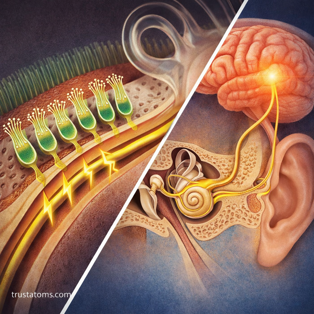
Sound transduction occurs when movement of the basilar membrane causes hair cell stereocilia to bend relative to surrounding structures. This mechanical displacement opens specialized ion channels, allowing positively charged ions to enter the hair cells and change their electrical potential.
Inner hair cells then release neurotransmitter onto auditory nerve endings. These nerve fibers generate action potentials that carry sound information toward the brain. Outer hair cells help amplify and sharpen cochlear vibrations, improving sensitivity and frequency discrimination.
This is an example of the processes described in sensory receptors and transduction mechanisms, where specialized cells transform environmental stimuli into biological signals.
Once activated, auditory nerve fibers transmit information through interconnected brain regions. These connections are part of the broader neural pathways in sensory processing.
The timing and distribution of nerve impulses help represent sound properties. Understanding neural coding in sensory systems explains how patterns of electrical activity convey information about frequency, intensity, and timing.
How the Cochlea Detects Different Pitches
Sound frequency, measured in hertz (Hz), strongly influences perceived pitch. Higher-frequency sounds generally produce higher pitches, while lower-frequency sounds produce lower pitches.
The cochlea separates frequencies through a spatial arrangement called tonotopic organization:
- High frequencies: Produce their strongest basilar membrane responses near the stiff base of the cochlea.
- Low frequencies: Produce their strongest responses toward the more flexible cochlear apex.
This frequency map is preserved through much of the auditory nervous system, allowing the brain to distinguish different components of complex sounds.
An Everyday Example: Hearing a 1,000 Hz Tone
Imagine hearing a steady tone at 1,000 Hz. Its sound wave produces 1,000 pressure oscillations per second. These oscillations vibrate the eardrum and ossicles, generating fluid movement inside the cochlea.
A particular region of the basilar membrane responds strongly to this frequency, stimulating nearby hair cells. Auditory nerve activity then carries frequency and timing information to the brain, where the tone is perceived.
If the same tone becomes louder, the cochlear response generally increases and additional auditory nerve activity helps represent the greater intensity. Frequency and loudness are related to different properties of the sound wave.
How Auditory Signals Reach the Brain
Auditory nerve fibers form the cochlear division of cranial nerve VIII, the vestibulocochlear nerve. Signals enter the brainstem and travel through several processing centers before reaching the auditory cortex.
Major structures include the cochlear nuclei, superior olivary complex, inferior colliculus, and medial geniculate nucleus of the thalamus. Auditory information reaches both sides of the brain through interconnected pathways.
The auditory cortex, located primarily in the temporal lobe, contributes to identifying pitch, timing, and complex sound patterns. Additional brain networks support speech comprehension, sound recognition, and spatial hearing.
How the Brain Identifies Sound Direction
The brain estimates sound location by comparing information from both ears. Two useful cues are differences in the time a sound reaches each ear and differences in sound intensity between the ears.
The shape of the outer ear also changes incoming sound depending on its direction, providing information about whether a sound originates above, below, or behind the listener.
Natural Protection Against Sound Damage
Several structures help protect normal ear function:
- Earwax: Helps trap particles and protects the skin lining the ear canal.
- Middle ear muscle reflex: Reduces transmission of some sustained loud sounds but offers limited protection against sudden impulses.
- Eustachian tube: Helps maintain appropriate middle ear pressure for effective eardrum movement.
These mechanisms cannot reliably prevent noise-induced hearing damage. Limiting exposure to excessively loud sound and using suitable hearing protection remain important preventive measures.
Common Causes of Hearing Loss
Hearing loss is commonly classified according to the location of the underlying problem.
| Type | Affected Structures | Examples |
|---|---|---|
| Conductive | Outer or middle ear | Earwax blockage, middle ear fluid, eardrum damage |
| Sensorineural | Cochlea or auditory nerve | Age-related changes, noise injury, inner ear disorders |
| Mixed | Both conductive and sensorineural components | Combined middle ear and inner ear problems |
Tinnitus, the perception of ringing or other sounds without a corresponding external source, can occur alongside hearing disorders. Sudden unexplained hearing loss requires urgent medical assessment because timely treatment may affect the outcome.
Frequently Asked Questions
What part of the ear converts sound into electrical signals?
Sensory hair cells within the organ of Corti in the cochlea convert mechanical movement into electrical changes. Inner hair cells communicate with auditory nerve fibers through neurotransmitter release.
What is the difference between pitch and loudness?
Pitch is primarily associated with sound frequency, while loudness is a perceptual response strongly influenced by sound intensity. Both properties are represented through patterns of activity in the auditory system.
Can damaged inner ear hair cells grow back?
In humans, damaged cochlear hair cells generally do not regenerate naturally. Permanent hair cell loss can therefore cause lasting hearing impairment, making prevention of excessive noise exposure particularly important.
Hearing depends on a precise sequence that transforms airborne pressure waves into mechanical vibrations, hair cell responses, and neural signals. The cochlea separates sound frequencies while auditory pathways and brain networks interpret timing, intensity, and meaning. Understanding these processes explains both normal hearing and how damage to individual structures can interfere with sound perception.
This article is for general education and is not medical advice. Talk to a qualified health professional about any medical concern.
Sources: Guyton and Hall Textbook of Medical Physiology; Principles of Neural Science; National Institute on Deafness and Other Communication Disorders (NIDCD).