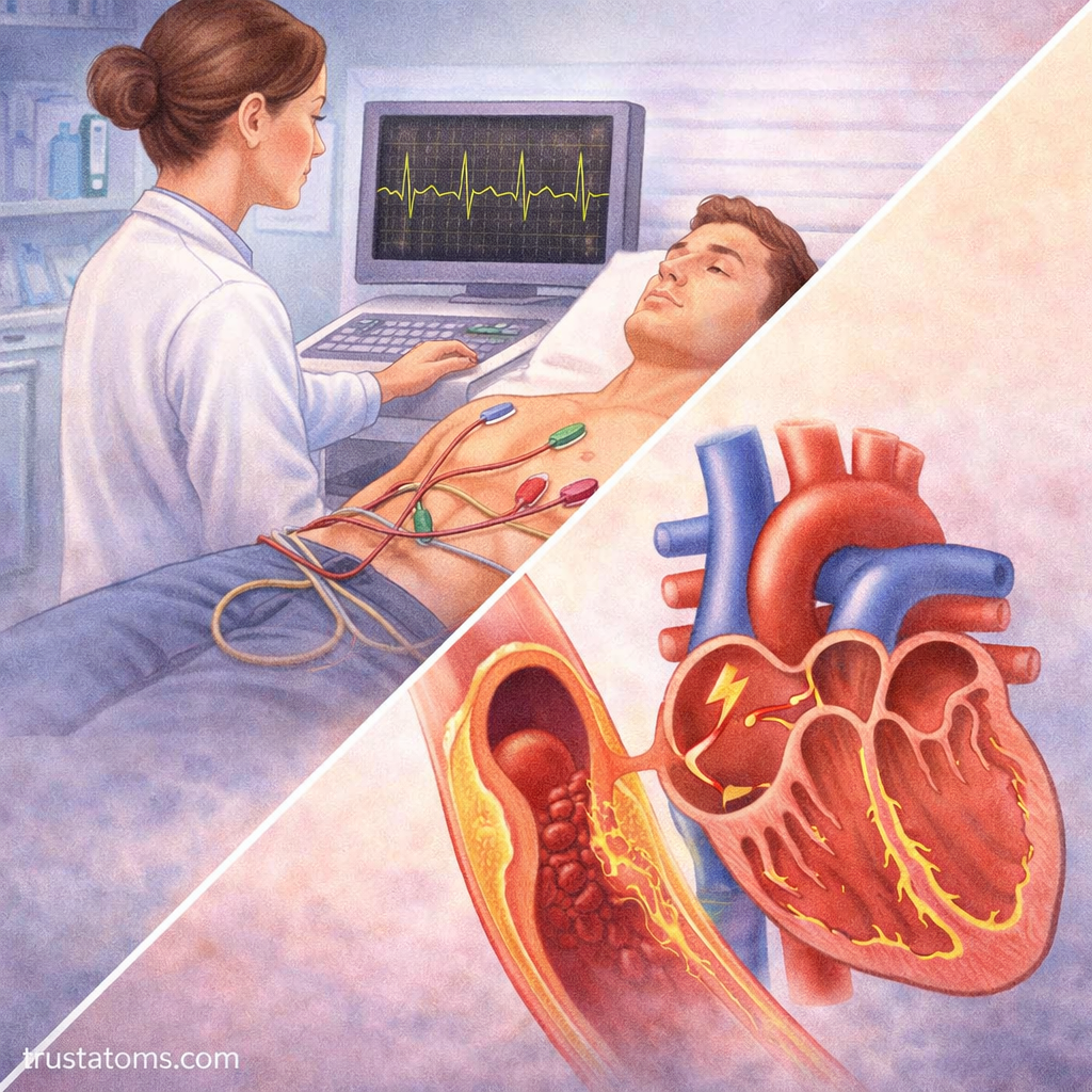
An electrocardiogram (ECG or EKG) is a simple, non-invasive test that records the electrical activity of the heart. It provides valuable insight into how the heart beats, how signals travel through it, and whether the rhythm is normal.
Understanding the electrical conduction pathways of the heart is essential for interpreting ECG readings and recognizing how the heart maintains a steady, coordinated rhythm.
What Is an Electrocardiogram (ECG)?
An ECG measures the electrical signals that trigger each heartbeat. These signals cause the heart muscles to contract and pump blood throughout the body.
What an ECG Shows
- Heart rate
- Heart rhythm
- Timing of electrical impulses
- Signs of abnormalities or damage
Electrodes placed on the skin detect these electrical changes and display them as waveforms.
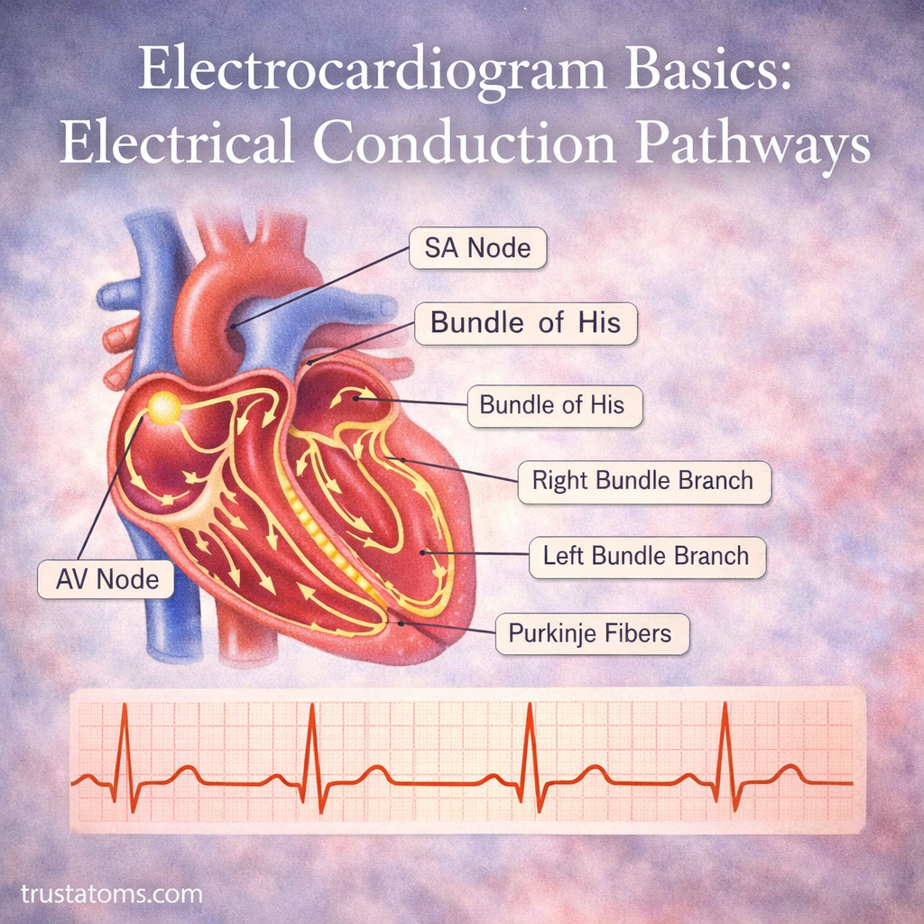
The Heart’s Electrical Conduction System
The heart has a built-in electrical system that controls the sequence of contractions.
Key Components
- Sinoatrial (SA) node
- Atrioventricular (AV) node
- Bundle of His
- Right and left bundle branches
- Purkinje fibers
Each part plays a specific role in coordinating the heartbeat.
Step-by-Step Electrical Pathway
The conduction pathway ensures that the heart beats in a synchronized and efficient manner.
1. Sinoatrial (SA) Node – The Natural Pacemaker
Located in the right atrium, the SA node initiates the electrical signal.
- Sets the heart rate
- Generates impulses automatically
- Causes the atria to contract
2. Atrial Conduction
The electrical signal spreads across both atria.
- Atria contract
- Blood is pushed into the ventricles
3. Atrioventricular (AV) Node – The Delay Center
The AV node slows the electrical signal before it reaches the ventricles.
- Allows ventricles time to fill with blood
- Ensures proper timing between atrial and ventricular contraction
4. Bundle of His and Bundle Branches
The signal travels from the AV node into the Bundle of His and then divides into:
- Right bundle branch
- Left bundle branch
These pathways carry the signal toward the ventricles.
5. Purkinje Fibers
Purkinje fibers distribute the electrical signal throughout the ventricles.
- Ventricles contract
- Blood is pumped to the lungs and the rest of the body
ECG Waves and What They Represent
An ECG trace consists of several waves, each corresponding to a specific electrical event.
P Wave
- Represents atrial depolarization
- Indicates atrial contraction
QRS Complex
- Represents ventricular depolarization
- Signals ventricular contraction
T Wave
- Represents ventricular repolarization
- Indicates recovery phase before the next beat
Why Electrical Conduction Matters
The heart’s ability to pump effectively depends on proper electrical coordination.
Key Functions
- Maintains a consistent heart rhythm
- Ensures efficient blood flow
- Prevents irregular contractions
Disruptions in conduction can lead to arrhythmias and other cardiac issues.
Common Conduction Abnormalities
Problems in the electrical system can affect heart function.
Arrhythmias
Irregular heart rhythms caused by abnormal electrical activity.
Examples:
- Tachycardia (fast heart rate)
- Bradycardia (slow heart rate)
Heart Block
Occurs when electrical signals are delayed or blocked.
- First-degree (mild delay)
- Second-degree (intermittent block)
- Third-degree (complete block)
Atrial Fibrillation
- Irregular and rapid atrial activity
- Reduces efficient blood flow
How an ECG Is Performed
An ECG is quick, painless, and widely used in medical settings.
Procedure Overview
- Electrodes are placed on the chest, arms, and legs
- Electrical signals are recorded
- A waveform is generated on a monitor or paper
The test typically takes only a few minutes.
Factors That Affect ECG Readings
Several factors can influence ECG results.
Physiological Factors
- Heart rate
- Age
- Physical fitness
External Factors
- Electrode placement
- Movement during testing
- Electrical interference
Accurate readings depend on proper technique and conditions.
Importance in Medical Diagnosis
ECGs are essential tools for diagnosing and monitoring heart conditions.
What They Can Detect
- Arrhythmias
- Heart attacks (myocardial infarction)
- Conduction delays
- Electrolyte imbalances
They are often used in emergency and routine care.
Tips for Understanding ECG Basics
While ECG interpretation can be complex, understanding the basics helps build a foundation.
Key Takeaways
- The heart’s rhythm is controlled by electrical signals
- Each ECG wave corresponds to a specific event
- Proper conduction ensures efficient heart function
Final Thoughts
The electrical conduction pathways of the heart are essential for maintaining a steady and coordinated heartbeat. Through the SA node, AV node, and specialized conduction fibers, the heart ensures that each contraction occurs in the correct sequence.
An electrocardiogram provides a window into this system, allowing healthcare providers to assess heart health and detect abnormalities. Understanding these basics makes it easier to appreciate how the heart functions and why electrical balance is so important.