The human heart beats with remarkable precision thanks to an internal electrical system known as the cardiac conduction system. This system coordinates each heartbeat, ensuring blood is pumped efficiently throughout the body.
Understanding how electrical impulses travel from the sinoatrial (SA) node to the Purkinje fibers provides insight into how the heart maintains rhythm—and what happens when that rhythm is disrupted.
What Is the Cardiac Conduction System?
The cardiac conduction system is a network of specialized cells that generate and transmit electrical impulses. These impulses trigger the heart muscle (myocardium) to contract in a coordinated way.
Instead of relying on the brain for every beat, the heart has its own built-in pacemaker system that controls timing and rhythm automatically.
Key functions include:
- Initiating the heartbeat
- Coordinating atrial and ventricular contractions
- Maintaining a consistent heart rate
- Adapting to the body’s demands (e.g., exercise or rest)
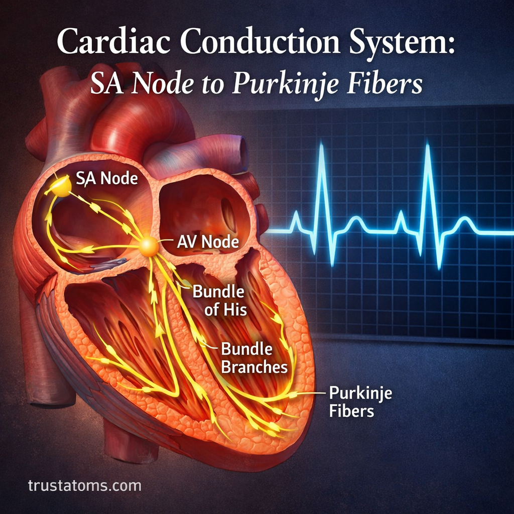
Overview of the Electrical Pathway
The conduction pathway follows a specific sequence:
- SA Node (pacemaker)
- Atrial pathways
- AV Node (delay center)
- Bundle of His
- Bundle branches
- Purkinje fibers
This sequence ensures blood flows efficiently from the atria to the ventricles and then out to the body.
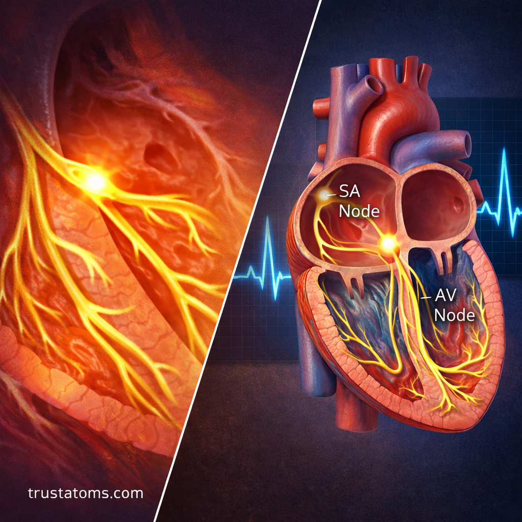
SA Node: The Heart’s Natural Pacemaker
The sinoatrial (SA) node is located in the right atrium near the opening of the superior vena cava.
Key Roles of the SA Node
- Generates electrical impulses automatically
- Sets the heart rate (typically 60–100 beats per minute at rest)
- Initiates atrial contraction
Because of its dominant role, the SA node is often referred to as the heart’s natural pacemaker.
Why It Matters
If the SA node fails or becomes impaired, other parts of the conduction system can take over—but usually at a slower rate.
Atrial Conduction Pathways
Once the SA node fires, the electrical impulse spreads across both atria through specialized conduction pathways and muscle fibers.
What Happens During This Phase
- Both atria contract (atrial systole)
- Blood is pushed into the ventricles
- Electrical signal moves toward the AV node
This step ensures the ventricles are properly filled before they contract.
AV Node: The Critical Delay Point
The atrioventricular (AV) node is located at the junction between the atria and ventricles.
Main Functions
- Receives the electrical signal from the atria
- Delays the signal briefly (about 0.1 seconds)
- Allows ventricles time to fill with blood
Why the Delay Is Essential
Without this pause:
- The atria and ventricles would contract simultaneously
- Ventricular filling would be incomplete
- Cardiac efficiency would drop significantly
The AV node acts as a gatekeeper, ensuring proper timing between upper and lower chambers.
Bundle of His: The Electrical Bridge
After passing through the AV node, the impulse enters the Bundle of His, located in the interventricular septum.
Key Characteristics
- Only electrical connection between atria and ventricles
- Rapidly transmits impulses downward
- Splits into left and right bundle branches
This structure ensures the signal moves efficiently into the ventricles.
Right and Left Bundle Branches
The Bundle of His divides into:
- Right bundle branch (serves the right ventricle)
- Left bundle branch (serves the left ventricle)
Their Role
- Carry electrical impulses along the interventricular septum
- Direct signals toward the apex of the heart
- Prepare the ventricles for coordinated contraction
The left bundle branch is thicker because the left ventricle requires more force to pump blood throughout the body.
Purkinje Fibers: Rapid Signal Distribution
Purkinje fibers are a network of specialized fibers that spread throughout the ventricular walls.
Key Functions
- Distribute electrical impulses rapidly across ventricles
- Trigger ventricular contraction (ventricular systole)
- Ensure synchronized contraction from the apex upward
Why This Matters
This upward contraction pattern:
- Maximizes blood ejection
- Improves pumping efficiency
- Maintains proper circulation
Purkinje fibers conduct impulses faster than any other part of the system, allowing near-simultaneous activation of ventricular muscle cells.
Step-by-Step Summary of the Conduction Process
Here’s a simplified sequence of how one heartbeat occurs:
- SA node generates an impulse
- Atria contract and push blood into ventricles
- AV node delays the signal
- Signal travels through Bundle of His
- Bundle branches carry signal to ventricles
- Purkinje fibers distribute impulse
- Ventricles contract and pump blood out
This entire process happens in less than a second and repeats continuously.
How the System Maintains Rhythm
The conduction system is influenced by the autonomic nervous system:
Sympathetic Nervous System
- Increases heart rate
- Speeds up conduction
- Active during stress or exercise
Parasympathetic Nervous System
- Slows heart rate
- Reduces conduction speed
- Dominant during rest
This balance allows the heart to adapt instantly to changing conditions.
Common Issues in the Conduction System
Disruptions in this system can lead to abnormal heart rhythms (arrhythmias).
Examples Include
- Bradycardia (slow heart rate)
- Tachycardia (fast heart rate)
- Heart block (impaired AV conduction)
- Atrial fibrillation (irregular atrial activity)
Causes May Include
- Aging or degeneration of conduction tissue
- Ischemia (reduced blood supply)
- Electrolyte imbalances
- Medications affecting electrical activity
Understanding the pathway helps identify where these issues originate.
Why the Cardiac Conduction System Is Essential
The heart’s ability to function independently and rhythmically depends entirely on this electrical system.
Without it:
- Blood circulation would become inefficient
- Oxygen delivery would decrease
- Organs and tissues would fail to function properly
The precision and speed of this system are what make continuous life-sustaining circulation possible.
Final Thoughts
The cardiac conduction system is a finely tuned electrical network that ensures every heartbeat is properly timed and effective. From the SA node initiating the signal to the Purkinje fibers triggering powerful ventricular contractions, each component plays a vital role.
By understanding this pathway, it becomes easier to appreciate how the heart works—and how disruptions can impact overall health.