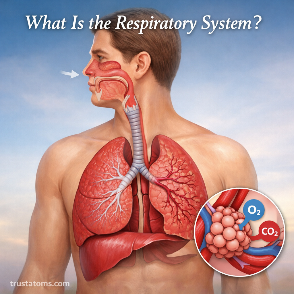
The respiratory system is the body’s network of organs and tissues responsible for breathing and exchanging gases with the environment. Its main job is to bring oxygen into the bloodstream and remove carbon dioxide produced by the body’s cells.
In human anatomy, the respiratory system includes the nose, throat, airways, lungs, and muscles that help move air. These structures work with the circulatory system to deliver oxygen to tissues and carry carbon dioxide back to the lungs.
Breathing usually happens automatically, whether you are sleeping, studying, or exercising. Inside the lungs, millions of tiny air sacs allow gases to move between inhaled air and blood, supporting cellular energy production and the regulation of blood acidity.
Overview of the Respiratory System
The respiratory system has two closely connected tasks: ventilation, which moves air into and out of the lungs, and gas exchange, which transfers oxygen and carbon dioxide between air and blood. Ventilation supplies fresh air to the lungs, while gas exchange allows the blood to collect oxygen and release carbon dioxide.
The route air follows is organized into a branching network of passages. Airway Anatomy explains how these passages connect the nose and mouth to the microscopic air sacs inside the lungs.
The efficiency of gas transfer depends partly on the condition of the alveoli and their surrounding capillaries. Diffusion Capacity in Lung Function examines how effectively gases move across the lung’s exchange surface.
Five Main Functions of the Respiratory System
- Oxygen uptake: Transfers oxygen from inhaled air into pulmonary capillary blood.
- Carbon dioxide removal: Eliminates carbon dioxide generated during cellular metabolism.
- Blood pH regulation: Helps control acidity by adjusting carbon dioxide removal.
- Voice production: Provides airflow that passes through the vocal folds to produce sound.
- Smell and airway protection: Supports odor detection and helps filter, warm, and humidify incoming air.
Major Parts of the Respiratory System
Each part of the respiratory system has a specific role in breathing and gas exchange. The conducting airways transport air, while the respiratory portions of the lungs provide surfaces where gases can diffuse.
| Structure | Main Function |
|---|---|
| Nose and nasal cavity | Filter, warm, and humidify incoming air |
| Pharynx | Conduct air from the nose or mouth toward the larynx |
| Larynx | Protect the airway and produce sound |
| Trachea | Carry air toward the bronchi |
| Bronchi | Distribute air into the lungs |
| Bronchioles | Regulate and distribute airflow through smaller passages |
| Alveoli | Exchange oxygen and carbon dioxide with blood |
| Diaphragm | Generate pressure changes needed for ventilation |
Upper Respiratory Tract
The upper respiratory tract prepares air before it reaches the lungs. It includes the nose, nasal cavity, and pharynx in a common anatomical classification.
The nasal passages contain mucus and tiny hairlike structures called cilia that help trap and move particles. Blood vessels near the nasal lining warm incoming air, while moisture from the lining helps prevent excessive drying of deeper airways.
The paranasal sinuses are air-filled spaces connected to the nasal cavity. They produce mucus and contribute to voice resonance, although their precise physiological functions are still studied.
Lower Respiratory Tract
The lower respiratory tract generally includes the larynx, trachea, bronchi, bronchioles, and lungs. The larynx contains the vocal folds, while the trachea is supported by cartilage that helps keep its airway open.
The trachea divides into two main bronchi, one entering each lung. These branches repeatedly divide into smaller bronchi and bronchioles. At the ends of the respiratory airways are alveoli, where most gas exchange occurs.
How the Respiratory System Works During Breathing
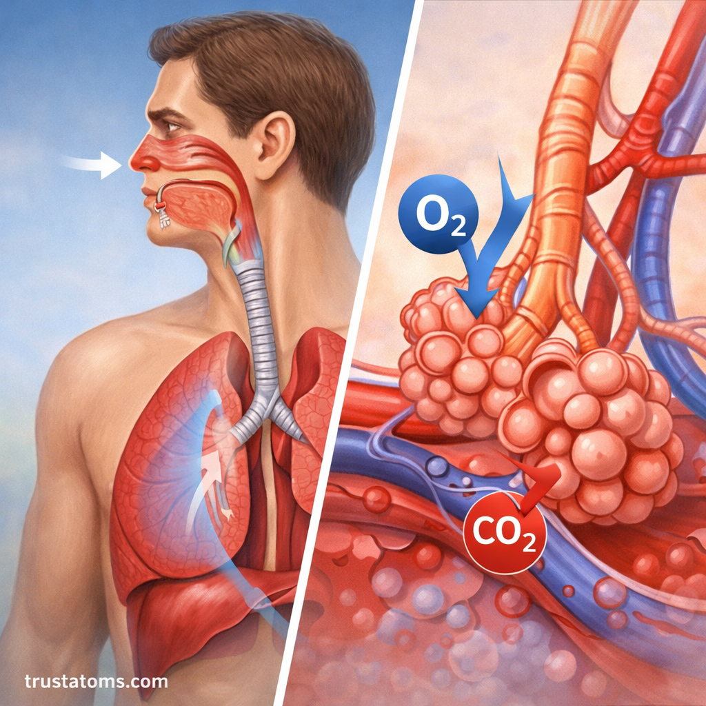
Breathing depends on pressure differences between the atmosphere and the air inside the lungs. Changes in chest volume create these differences, causing air to flow into or out of the respiratory passages.
Inhalation: How Air Enters the Lungs
- The diaphragm contracts and moves downward, becoming flatter.
- The external intercostal muscles help lift and expand the rib cage.
- The chest cavity expands, allowing the lungs to expand with it.
- Pressure inside the alveoli briefly falls below atmospheric pressure, so air flows inward.
Air passes through the conducting airways before reaching the respiratory regions of the lungs. The diaphragm is the main muscle of quiet inhalation, although additional muscles can assist during strenuous activity.
Exhalation: How Air Leaves the Lungs
During relaxed exhalation, the diaphragm and inspiratory muscles relax. Elastic recoil of the lungs and chest wall reduces lung volume, briefly raising alveolar pressure above atmospheric pressure and driving air outward.
Forceful exhalation, such as during vigorous exercise or coughing, recruits additional muscles, including abdominal and internal intercostal muscles.
How Oxygen and Carbon Dioxide Move Through the Alveoli
Alveoli are microscopic air sacs surrounded by networks of pulmonary capillaries. Their thin walls provide a short diffusion distance between inhaled air and circulating blood.
Oxygen moves from alveolar air, where its partial pressure is higher, into blood arriving from the pulmonary arteries. Carbon dioxide moves in the opposite direction because its partial pressure is generally higher in the incoming blood than in alveolar air.
These movements occur through diffusion, meaning gases move down their partial-pressure gradients without requiring a pump to push individual gas molecules across the membrane.
How Blood Transports Respiratory Gases
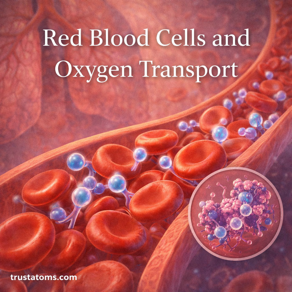
Most oxygen in the blood binds reversibly to hemoglobin, a protein inside red blood cells. A smaller amount remains dissolved in plasma. Hemoglobin releases oxygen in tissues where conditions favor unloading.
Carbon dioxide is transported in several forms, primarily as bicarbonate ions, with smaller amounts bound to proteins or dissolved in blood. The lungs eliminate carbon dioxide when it diffuses into alveoli and is exhaled.
Worked Example: How Much Air Do You Breathe in One Minute?
A useful way to understand ventilation is to calculate minute ventilation, the total volume of air entering or leaving the lungs each minute.
Consider a resting adult who breathes 12 times per minute, with approximately 500 milliliters of air moving in or out during each normal breath. These are common textbook example values, not fixed measurements for every person.
- Breathing rate: 12 breaths per minute.
- Tidal volume: 500 milliliters, or 0.5 liters, per breath.
- Calculation: 12 breaths per minute × 0.5 liters per breath = 6 liters per minute.
In this example, minute ventilation is 6 liters of air per minute. However, not all inhaled air reaches gas-exchanging alveoli. Some remains in the conducting airways, which are part of anatomical dead space.
If anatomical dead space is approximately 150 milliliters per breath, the estimated alveolar ventilation would be (500 − 150) × 12 = 4,200 milliliters, or 4.2 liters per minute. This simplified calculation assumes anatomical dead space approximates physiological dead space in a healthy resting adult.
How Breathing Helps Regulate Blood pH
Carbon dioxide is more than a waste gas. In blood, it participates in chemical reactions that produce carbonic acid and bicarbonate, influencing hydrogen ion concentration and therefore pH.
When ventilation increases relative to carbon dioxide production, blood carbon dioxide levels tend to fall, which can raise blood pH. When ventilation decreases, carbon dioxide may accumulate and lower pH.
The respiratory system works with the kidneys and chemical buffer systems to maintain acid-base balance. Breathing can adjust carbon dioxide relatively quickly, while kidney responses generally develop over a longer period.
How the Respiratory System Works With Other Body Systems
Circulatory System
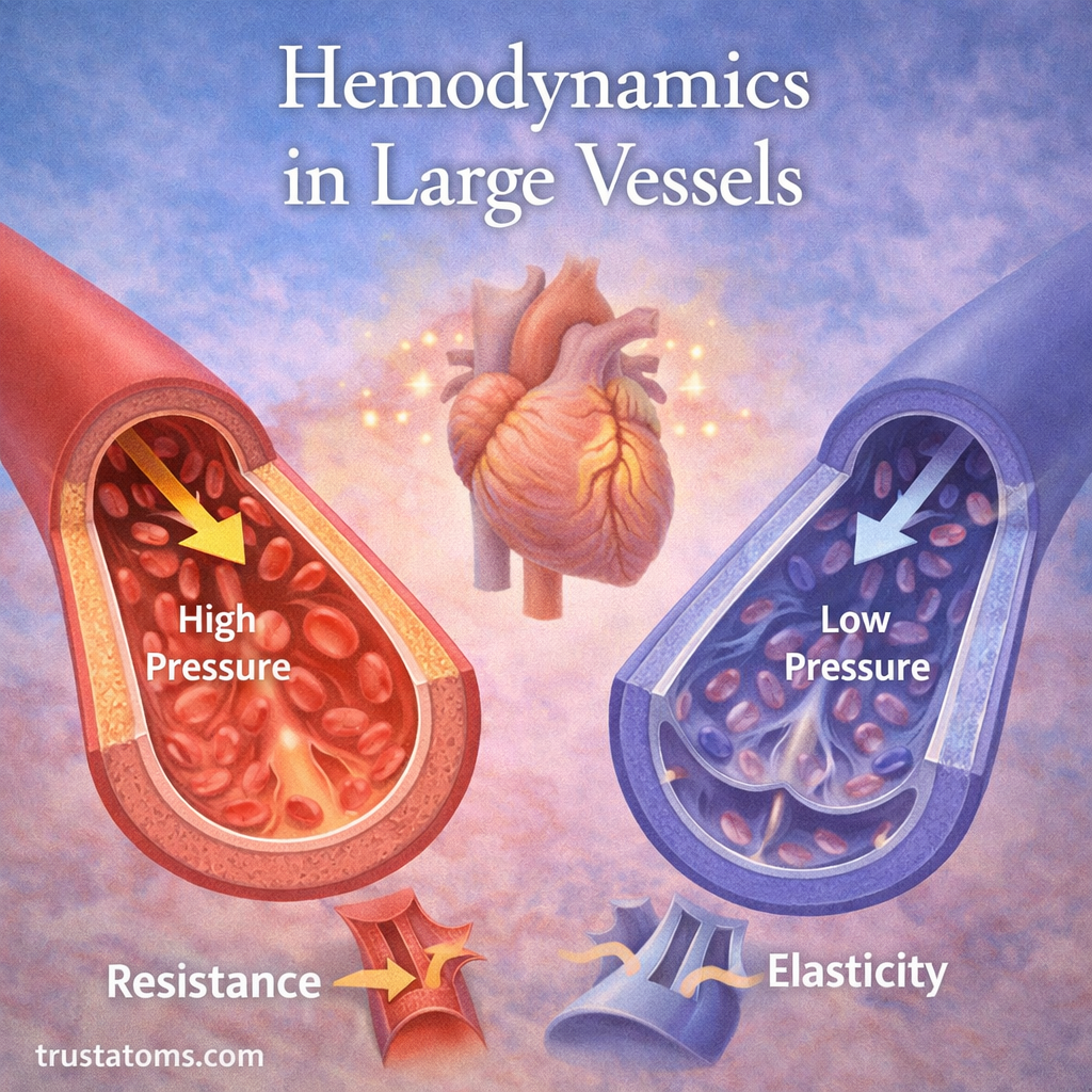
The pulmonary circulation carries blood from the right side of the heart to the lungs, where gas exchange occurs. Oxygenated blood returns to the left side of the heart and is pumped to tissues throughout the body.
Nervous System
Breathing rhythms are generated by neural circuits in the brainstem. Chemoreceptors detect changes related to carbon dioxide, pH, and oxygen, helping adjust breathing depth and frequency as the body’s needs change.
Muscular System
The diaphragm and intercostal muscles generate the mechanical changes needed for ventilation. During exercise, respiratory muscles work harder to support increased airflow and the removal of additional carbon dioxide.
Common Respiratory Conditions
Respiratory disorders can affect airway diameter, lung tissue, gas exchange, or breathing mechanics. Their symptoms and underlying causes differ.
- Asthma: A condition involving variable airway narrowing and inflammation, often causing wheezing, coughing, or shortness of breath.
- Bronchitis: Inflammation of the bronchi that can cause coughing and mucus production.
- Pneumonia: Infection and inflammation of lung tissue that may interfere with gas exchange.
- Chronic obstructive pulmonary disease (COPD): A group of conditions characterized by persistent airflow limitation, commonly associated with smoking or other harmful exposures.
- Pulmonary fibrosis: Scarring of lung tissue that can make the lungs less flexible and impair oxygen transfer.
Severe breathing difficulty, blue or gray discoloration of the lips, new confusion, or chest pain accompanied by shortness of breath requires urgent medical evaluation.
How to Support Respiratory Health
Respiratory health depends on healthy airways, functioning lung tissue, effective breathing muscles, and adequate protection against harmful exposures.
- Avoid tobacco smoke: Smoking damages airways and lung tissue, while secondhand smoke also creates health risks.
- Reduce pollutant exposure: Improve ventilation where appropriate and limit exposure to smoke, dust, and harmful fumes.
- Stay physically active: Regular activity improves cardiovascular and muscular conditioning, helping the body use oxygen efficiently.
- Prevent infections: Recommended vaccinations, handwashing, and other appropriate precautions reduce risks from respiratory infections.
- Manage existing conditions: Follow professional guidance for asthma, COPD, or other diagnosed respiratory disorders.
Common Misconceptions
Misconception: The Lungs Pull Air In by Themselves
The lungs do not contain muscles that actively expand them during normal breathing. The diaphragm and other respiratory muscles change chest volume, creating pressure differences that move air.
Misconception: Exhaled Air Contains No Oxygen
Exhaled air still contains substantial oxygen. At sea level, ordinary atmospheric air contains about 21% oxygen, while mixed exhaled air commonly contains approximately 16%, depending on breathing conditions and activity.
Misconception: Breathing Faster Always Improves Oxygen Delivery
Faster breathing does not necessarily improve oxygen delivery. Effective gas exchange depends on adequate alveolar ventilation, blood flow, and lung function, and excessive ventilation can lower carbon dioxide unnecessarily.
Misconception: Oxygen Is the Main Trigger for Normal Breathing
In healthy people under ordinary conditions, changes in carbon dioxide and the associated changes in pH provide a major drive for breathing regulation. Low oxygen can also stimulate breathing, especially when oxygen levels fall substantially.
Frequently Asked Questions
What Are the Main Organs of the Respiratory System?
The main structures include the nose, nasal cavity, pharynx, larynx, trachea, bronchi, bronchioles, and lungs. The diaphragm and other respiratory muscles help move air through these passages.
What Is the Main Function of the Respiratory System?
Its primary function is to bring oxygen into the bloodstream and remove carbon dioxide. It also helps regulate blood pH, supports speech, and contributes to smell and airway protection.
Where Does Gas Exchange Occur in the Lungs?
Most gas exchange occurs across the thin walls of alveoli and surrounding pulmonary capillaries. Oxygen diffuses into blood, while carbon dioxide diffuses toward the alveolar air.
What Is the Difference Between Breathing and Cellular Respiration?
Breathing is the physical movement of air into and out of the lungs. Cellular respiration refers to metabolic processes through which cells produce usable energy, with aerobic pathways using oxygen and producing carbon dioxide.
How Many Breaths Does an Adult Take Per Minute?
A typical resting respiratory rate for a healthy adult is approximately 12–20 breaths per minute. The rate varies with activity, age, temperature, health, and other factors.
The respiratory system combines mechanical ventilation with microscopic gas exchange to supply oxygen and remove carbon dioxide. Its airways, alveoli, breathing muscles, and blood vessels work together to support cellular metabolism, regulate blood chemistry, and respond to changing activity levels.
This article is for general education and is not medical advice. Talk to a qualified health professional about any medical concern.
Sources: OpenStax, Anatomy and Physiology 2e; National Heart, Lung, and Blood Institute (NHLBI); National Library of Medicine, MedlinePlus; West’s Respiratory Physiology: The Essentials.