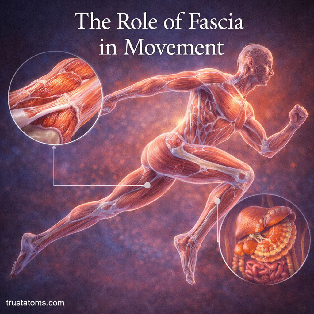
Fascia supports human movement by surrounding muscles, connecting tissues, transmitting mechanical forces, and allowing neighboring structures to slide against one another. Its collagen-rich layers contribute to stability and flexibility while working alongside muscles, tendons, bones, and joints to produce controlled motion.
When most people think about movement, they focus on muscles, bones, and joints. However, fascia adds another important dimension to human anatomy by connecting structures that might otherwise appear to function independently.
What Is Fascia?
Fascia is a connective tissue network that surrounds and links muscles, organs, and structures throughout the body. It includes sheets, bands, and layers of tissue that provide support while permitting movement between neighboring structures.
Fascia contains collagen fibers, varying amounts of elastin, specialized connective tissue cells called fibroblasts, and a water-rich extracellular matrix. Its composition and organization differ depending on the mechanical demands of each location.
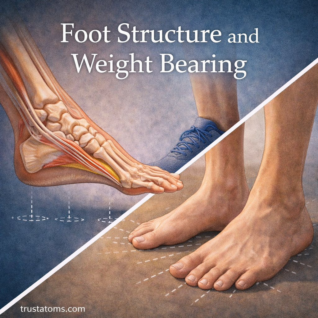
For example, the plantar fascia along the sole of the foot helps support the foot’s longitudinal arch and contributes to its mechanical behavior during walking. Its relationship with bones and joints is explored in Foot Structure and Weight Bearing.
Fascia is part of the broader Musculoskeletal System, where connective tissues help transfer forces between muscles and skeletal structures.
The Three Main Types of Fascia
Fascia is commonly described in three broad categories, although anatomical terminology and classification can vary.
Superficial Fascia
Superficial fascia lies beneath the skin and contains loose connective tissue and varying amounts of fat. It helps cushion underlying structures, provides pathways for blood vessels and nerves, and allows the skin to move over deeper tissues.
Deep Fascia
Deep fascia consists of denser connective tissue that surrounds muscles and separates muscle groups into compartments. It can form strong sheets, such as the fascia lata of the thigh, that contribute to structural support and mechanical force transmission.
Visceral Fascia
Visceral fascia surrounds and supports internal organs. Its connective tissue layers help maintain anatomical relationships while permitting the movement required for functions such as breathing and digestion.
How Fascia Transmits Force During Movement
Muscles generate force through contraction, but that force is not transmitted exclusively through tendons. Connective tissues surrounding muscle fibers and muscle groups can also transfer tension to adjacent structures.
This process is called myofascial force transmission. It means that mechanical forces can pass through connective tissue connections within and between muscles, although the amount transferred depends on anatomy, movement, and loading conditions.
Consider lifting a heavy grocery bag. Your hand and forearm muscles grip the handle, while muscles around the shoulder and trunk help stabilize your body. Fascial tissues contribute to the transfer and distribution of forces across these regions, but coordinated muscle contractions remain the primary drivers of movement.
Fascial Elasticity and Energy Storage
Some fascial structures stretch slightly under tension and recoil when that tension decreases. This elastic behavior can contribute to energy storage and return during repetitive movements.
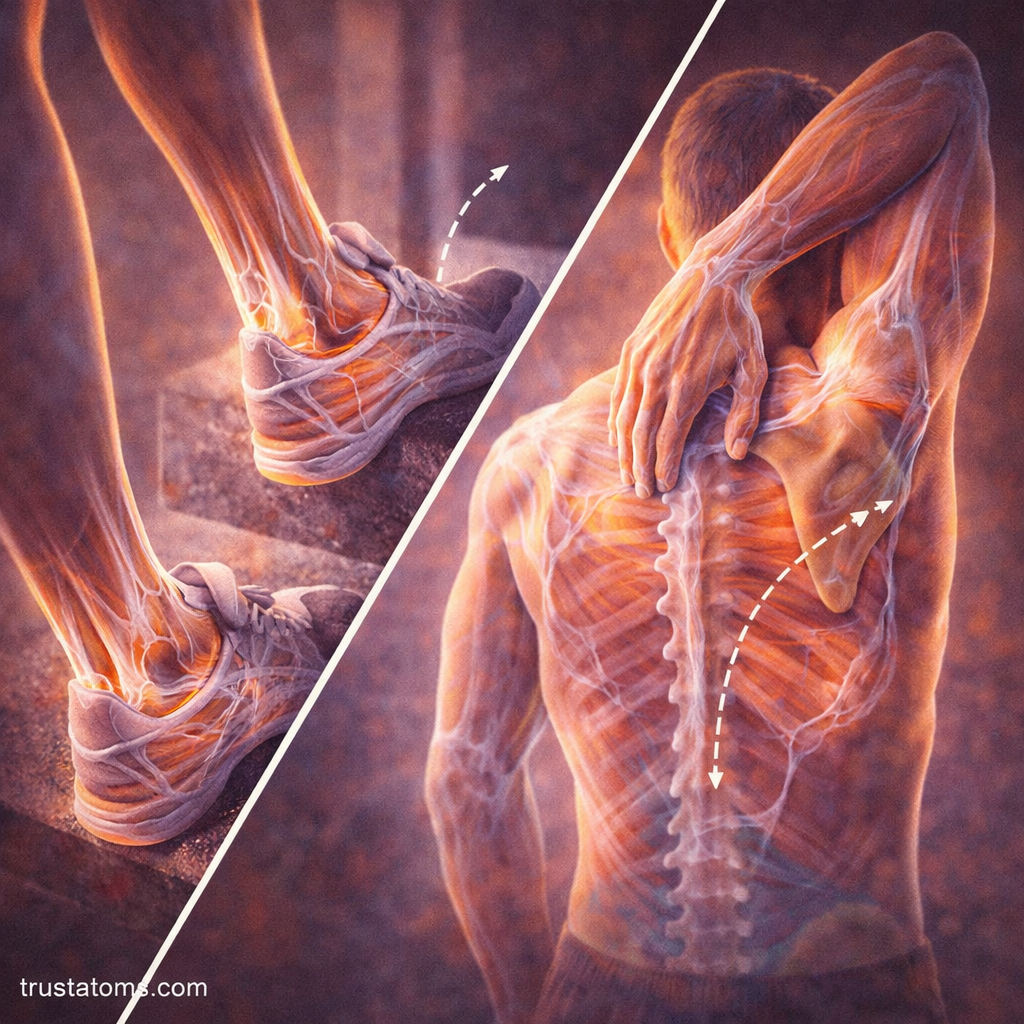
Walking is one example in which fascial elasticity supports efficient motion. As weight moves across the foot, the plantar fascia experiences tension and contributes to the mechanical support of the arch.
Fascia also exhibits viscoelastic behavior, meaning its response depends partly on how quickly and how long a force is applied. Under sustained loading, some tissues gradually deform, while under repeated loading they may show changes in stiffness.
Not all fascial tissues behave like powerful springs. The contribution of elastic energy depends on the tissue’s structure, location, and the movement being performed.
Fascia, Flexibility, and Range of Motion
Flexibility depends on several factors, including joint anatomy, muscle length, nervous system responses, and the mechanical properties of connective tissues.
Fascial layers can glide relative to neighboring tissues, helping muscles and other structures move without excessive friction. Loose connective tissue and substances such as hyaluronan contribute to this sliding behavior.
After injury, surgery, or prolonged immobilization, scar tissue and altered connective tissue organization can sometimes restrict movement. However, a feeling of tightness does not necessarily mean fascia is physically shortened or damaged.
Stretching may improve range of motion through a combination of changes in stretch tolerance and tissue behavior. The relative contribution of fascia varies, and improvements should not automatically be attributed to permanent fascial lengthening.
How Fascia Contributes to Posture and Stability
Deep fascial sheets provide structural connections between muscles and regions of the skeleton. These connections can help distribute tension and resist certain movements.
The thoracolumbar fascia, for example, forms a layered connective tissue structure in the lower back. It connects with several trunk and back muscles and contributes to the mechanical behavior of the lumbar region.
Posture is not maintained by fascia alone. Muscular activity, skeletal alignment, joint mechanics, and nervous system control work together to support standing and sitting positions.
There is also no single posture that guarantees freedom from discomfort. Regularly changing positions and maintaining sufficient strength and movement capacity are often more practical than trying to hold one supposedly perfect alignment.
Fascia and Sensory Feedback
Fascia contains sensory nerve endings, including receptors associated with mechanical stimulation and pain. This means fascial tissues can contribute information about tension, pressure, and tissue conditions.
Some fascial structures are richly innervated, while others contain fewer sensory endings. Their contribution to movement awareness depends on the tissue and its interaction with muscles, joints, and the nervous system.
Fascial tissues can also be sources of pain when irritated or injured. However, pain in a particular body region cannot reliably be attributed to fascia without considering other possible causes.
Fascia in Everyday Activities
Fascia experiences mechanical loading during nearly every physical activity, but different regions contribute in different ways.
- Walking: Plantar fascia helps support the arch as the foot accepts and transfers body weight.
- Running: Connective tissues experience repeated tension and contribute to the mechanical behavior of the limbs.
- Reaching: Fascial layers accommodate changes in muscle shape and position around the shoulder and trunk.
- Lifting: Deep fascia contributes to force transmission and structural support alongside active muscles.
How Movement and Exercise Affect Fascia
Like other connective tissues, fascia responds to mechanical loading over time. Regular activity provides physical stimulation that helps maintain normal tissue function, while prolonged immobilization can contribute to stiffness and reduced mobility.
Several practical habits support overall musculoskeletal health:
- Regular movement: Walking and varied daily activities expose tissues to different movement patterns.
- Progressive strengthening: Gradually increasing exercise demands allows muscles and connective tissues time to adapt.
- Mobility exercises: Comfortable stretching and controlled joint movements can help maintain functional range of motion.
- Adequate nutrition and hydration: Normal tissue maintenance requires sufficient nutrients and fluid balance.
- Foam rolling: May temporarily improve range of motion or reduce perceived soreness, although evidence does not establish that it permanently breaks apart fascial adhesions.
These approaches support movement without relying on claims that fascia must be manually released, detoxified, or reshaped to function normally.
Frequently Asked Questions
Is fascia the same as a tendon or ligament?
No. All three are connective tissues, but tendons primarily connect muscles to bones, ligaments typically connect bones to other bones, and fascia forms sheets and layers that surround, separate, and connect structures. Their compositions overlap, but their anatomical arrangements and functions differ.
Can fascia become tight or restricted?
Fascial tissues can develop changes in stiffness, sliding behavior, or organization, particularly after injury or immobilization. However, perceived tightness may also involve muscles, joints, or nervous system responses, so the sensation alone does not identify a fascial problem.
Does foam rolling permanently change fascia?
Foam rolling can produce short-term improvements in flexibility and perceived muscle soreness for some people. Current evidence does not clearly demonstrate that ordinary foam rolling permanently lengthens fascia or physically breaks down connective tissue adhesions.
Fascia contributes to movement through structural support, force transmission, tissue sliding, and sensory signaling. Its effects are closely connected to muscle activity and joint mechanics, making it one component of a coordinated movement system rather than an independent mechanism controlling the body.
This article is for general education and is not medical advice. Talk to a qualified health professional about any medical concern.
Sources: Gray’s Anatomy: The Anatomical Basis of Clinical Practice; Schleip and colleagues, Fascia: The Tensional Network of the Human Body; National Library of Medicine (NLM).