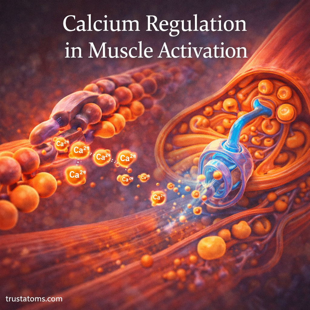
Calcium regulates muscle activation by acting as an internal signal that allows muscle proteins to generate force. When calcium ions (Ca²⁺) are released into a muscle cell, contraction can begin; when they are returned to storage, the muscle can relax.
Calcium plays a central role in how muscles contract and relax. While ATP provides the energy for movement, calcium ions control when the contractile proteins can interact. This relationship connects cellular physiology with human anatomy and explains how the nervous system produces controlled movements.
What Is Calcium’s Role in Muscles?
Inside skeletal muscle fibers, calcium acts as a signal that switches the contractile machinery between active and inactive states. A rapid increase in calcium concentration allows actin and myosin to interact, while a decrease helps stop contraction.
This process depends on changes in calcium concentration within the muscle cell, rather than simply the amount of calcium circulating in the bloodstream.
Blood calcium must also remain within a controlled range to support normal nerve and muscle function. Blood calcium homeostasis mechanisms explains how hormones, bones, kidneys, and the digestive system help regulate circulating calcium.
Where Is Calcium Stored Inside Muscle Cells?
Most calcium used to activate skeletal muscle contraction is stored in the sarcoplasmic reticulum (SR), a specialized membrane network surrounding the muscle’s contractile structures.
The SR contains calcium-binding proteins, including calsequestrin, that help it store substantial amounts of calcium. Specialized channels release calcium when the muscle receives an activating electrical signal.
At rest, calcium concentration in the cytosol, the fluid surrounding the muscle’s internal structures, remains low. This keeps the contractile proteins largely inactive until stimulation occurs.
How Muscle Activation Begins
Skeletal muscle activation begins when a motor neuron sends an electrical signal to a muscle fiber. The signal is converted into a change in calcium concentration through a process called excitation–contraction coupling.
The communication point between a motor neuron and skeletal muscle fiber is called the neuromuscular junction. Neuromuscular junction and muscle activation explains how the neurotransmitter acetylcholine initiates a muscle action potential.
The muscle fiber contains bundles of myofibrils made from repeating contractile units called sarcomeres. Skeletal muscle structure and function describes how these structures are organized to produce movement.
The Activation Sequence
- Nerve stimulation: A motor neuron releases acetylcholine at the neuromuscular junction.
- Electrical activation: The muscle fiber generates an action potential that spreads across its membrane and into transverse tubules, or T-tubules.
- Calcium release: Voltage-sensitive proteins in the T-tubules activate calcium-release channels in the sarcoplasmic reticulum.
- Protein activation: Released calcium binds to troponin, allowing actin and myosin to interact.
- Force production: Myosin uses energy from ATP to pull on actin filaments.
Calcium enables contraction, but it does not supply the energy for the movement. ATP use in muscle contraction explains how ATP powers cross-bridge cycling and supports calcium transport.
How Calcium Controls the Sliding Filament Mechanism
Actin and myosin are the main contractile proteins in skeletal muscle. Their interaction is regulated by two additional proteins: troponin and tropomyosin.
At rest, tropomyosin covers important myosin-binding sites on actin. When calcium binds to troponin C, the troponin complex changes shape and shifts tropomyosin away from these sites.
Myosin heads can then bind to actin and perform repeated force-generating cycles using ATP. The thin filaments slide past the thick filaments, shortening sarcomeres and producing muscle tension.
As long as sufficient calcium and ATP remain available, cross-bridge cycling can continue. The amount of force produced also depends on how many muscle fibers are activated and how frequently they receive stimulation.
How Calcium Reuptake Allows Muscle Relaxation
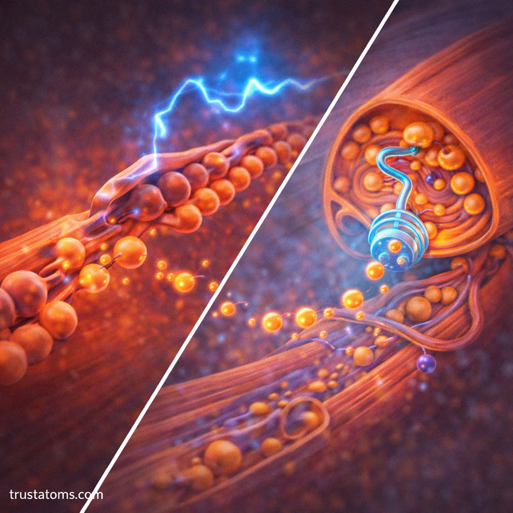
Relaxation begins when activating electrical signals stop and calcium-release channels close. Specialized pumps called sarcoplasmic/endoplasmic reticulum calcium ATPases (SERCA) transport calcium from the cytosol back into the sarcoplasmic reticulum.
SERCA pumps use ATP to move calcium against its concentration gradient. As cytosolic calcium decreases, calcium detaches from troponin, allowing tropomyosin to return toward its resting position and limit further cross-bridge formation.
Existing cross-bridges must also detach, which requires ATP binding to myosin. This is why ATP is needed for both contraction and normal relaxation.
How Repeated Calcium Signals Produce Stronger Contractions
A single muscle action potential usually produces a brief contraction called a twitch. If another signal arrives before the muscle has fully relaxed, calcium may remain elevated enough for the contractions to overlap.
This process, called temporal summation, can increase muscle tension. At sufficiently high stimulation frequencies, individual twitches merge into a sustained contraction called tetanus.
For example, holding a heavy grocery bag requires continued activation of muscle fibers rather than a single brief twitch. Repeated neural signals maintain the calcium activity needed for sustained force, while ATP supports cross-bridge cycling and calcium pumping.
How Calcium Regulation Differs Among Muscle Types
Calcium is essential for skeletal, cardiac, and smooth muscle contraction, but the activation mechanisms differ.
| Muscle type | How calcium activates contraction | Primary function |
|---|---|---|
| Skeletal muscle | Calcium released from the SR binds to troponin | Voluntary movement and posture |
| Cardiac muscle | Calcium entering the cell triggers additional SR release and binds to troponin | Rhythmic heart contractions |
| Smooth muscle | Calcium binds to calmodulin, activating enzymes that regulate myosin | Movement and tension in organs and blood vessels |
In cardiac muscle, calcium-induced calcium release helps coordinate contraction. Smooth muscle does not use the same troponin-based regulatory mechanism as skeletal muscle; instead, calcium–calmodulin signaling activates myosin light-chain kinase.
What Happens When Calcium Regulation Is Disrupted?
Normal muscle activity depends on properly timed calcium release, binding, and reuptake. Problems with these processes can interfere with force production or relaxation.
- Altered calcium release: Reduced release from the SR can contribute to weaker muscle contractions.
- Impaired calcium reuptake: Slower removal of calcium from the cytosol can delay relaxation.
- Muscle fatigue: Prolonged activity can alter calcium handling alongside energy availability and other cellular processes.
- Abnormal blood calcium: Significant changes in circulating calcium can affect nerve excitability and muscle function, sometimes causing spasms, weakness, or other symptoms.
These effects should not be confused with one another. Blood calcium disorders and disturbances in calcium signaling inside muscle cells involve related but distinct physiological mechanisms.
Frequently Asked Questions
Does calcium directly make muscles contract?
Calcium activates the contractile machinery but does not directly generate mechanical force. In skeletal muscle, it binds to troponin and permits actin–myosin interactions, while ATP supplies the energy for cross-bridge cycling.
Where does the calcium used in skeletal muscle contraction come from?
Most calcium that directly activates normal skeletal muscle contraction is released from the sarcoplasmic reticulum. Electrical signals traveling through T-tubules trigger this release.
Why is ATP needed for muscle relaxation?
ATP powers SERCA pumps that return calcium to the sarcoplasmic reticulum. ATP binding is also necessary for myosin to detach from actin, allowing the muscle to relax normally.
Calcium regulation connects electrical signals from motor neurons to the mechanical action of muscle fibers. Rapid calcium release activates contraction, while ATP-powered reuptake helps end it. The precise timing of these changes allows muscles to produce anything from brief, delicate movements to sustained force.
This article is for general education and is not medical advice. Talk to a qualified health professional about any medical concern.
Sources: Guyton and Hall Textbook of Medical Physiology; Boron and Boulpaep Medical Physiology; OpenStax Anatomy and Physiology 2e.