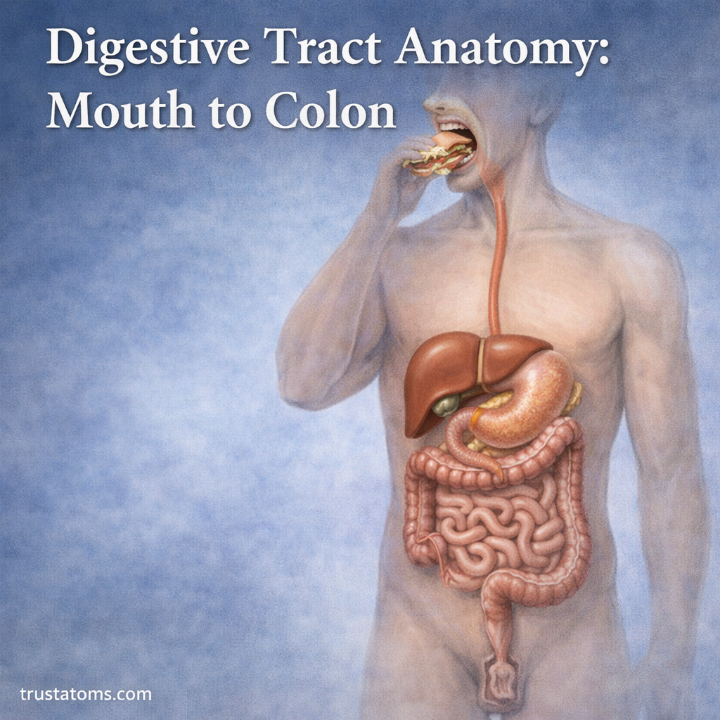
The digestive tract is a continuous muscular passage extending from the mouth through the pharynx, esophagus, stomach, small intestine, large intestine, rectum, and anus. Its organs work in sequence to break down food, absorb nutrients and water, and eliminate material the body cannot digest.
Each region has specialized tissues and movements suited to its function, making the digestive tract a useful example of how structure supports function in human anatomy. Understanding the route food follows also helps explain common problems such as acid reflux, nutrient malabsorption, and constipation.
Overview of the Digestive Tract
The digestive tract, also called the gastrointestinal or GI tract, forms a connected pathway through the body. Food moves through its organs in a generally one-way direction, undergoing mechanical breakdown, chemical digestion, absorption, and eventual elimination.
The tract is part of the larger digestive system, which also includes accessory organs such as the liver, gallbladder, and pancreas. For an introduction to these structures and their functions, see What Is the Digestive System?
The Mouth: Where Digestion Begins
Digestion starts when food enters the mouth. Teeth cut and grind food into smaller pieces, increasing the surface area available to digestive enzymes. Saliva moistens the food and contains salivary amylase, which begins breaking down starch.
The tongue moves food during chewing and shapes it into a soft mass called a bolus. Once swallowing begins, the bolus passes toward the pharynx.
Farther along the digestive tract, muscular valves regulate movement between different regions. These structures are explained in Sphincters and Flow Regulation.
Pharynx: The Swallowing Pathway
The pharynx is a muscular passage shared by the digestive and respiratory systems. During swallowing, coordinated muscle movements direct food toward the esophagus while protecting the airway.
The larynx moves upward and forward, the vocal folds close, and the epiglottis helps cover the laryngeal opening. These actions reduce the chance of food entering the trachea.
The sequence of voluntary and reflex actions involved is described in Swallowing Mechanics and Esophageal Transport.
Esophagus: Moving Food to the Stomach
The esophagus is a muscular tube that transports food to the stomach.
Wave-like muscular contractions called peristalsis propel swallowed food downward. This movement does not depend entirely on gravity, which is why swallowing can occur even when a person is lying down.
The lower esophageal sphincter relaxes to allow food into the stomach and normally helps prevent acidic stomach contents from flowing backward. When this protective barrier functions poorly, gastroesophageal reflux can occur.
Stomach: Mixing Food and Beginning Protein Digestion
The stomach is an expandable muscular organ located mainly in the upper abdomen. It temporarily stores swallowed food and mixes it with gastric secretions through coordinated contractions.
Gastric glands release hydrochloric acid and digestive enzymes. The acidic environment helps unfold proteins and activates pepsin, an enzyme that breaks proteins into smaller fragments. Mucus and bicarbonate help protect the stomach lining from its own secretions.
The resulting semi-liquid mixture is called chyme. The pyloric sphincter regulates its passage into the duodenum, the first section of the small intestine.
Small Intestine: The Main Site of Nutrient Absorption
The small intestine is the longest section of the digestive tract, measuring approximately 3–5 meters in many living adults, although measurements vary. It completes much of chemical digestion and absorbs most dietary nutrients.
It has three major regions:
- Duodenum: Receives chyme, bile, and pancreatic secretions that help digest carbohydrates, proteins, and fats.
- Jejunum: Absorbs a substantial proportion of digested nutrients, including sugars and amino acids.
- Ileum: Continues nutrient absorption and is especially important for absorbing vitamin B12 bound to intrinsic factor and recycling bile salts.
The intestinal lining contains folds, finger-like villi, and microscopic microvilli that greatly increase its absorptive surface. Blood capillaries carry many absorbed nutrients toward the liver, while lymphatic vessels transport much of the absorbed long-chain dietary fat.
Accessory Organs That Support Intestinal Digestion
Several organs contribute digestive substances without forming part of the passage food travels through.
- Liver: Produces bile, whose bile salts help emulsify dietary fats and support their absorption.
- Gallbladder: Stores and concentrates bile, releasing it into the small intestine when needed.
- Pancreas: Releases digestive enzymes and bicarbonate into the duodenum to break down nutrients and neutralize acidic chyme.
Large Intestine (Colon): Water Absorption and Waste Formation
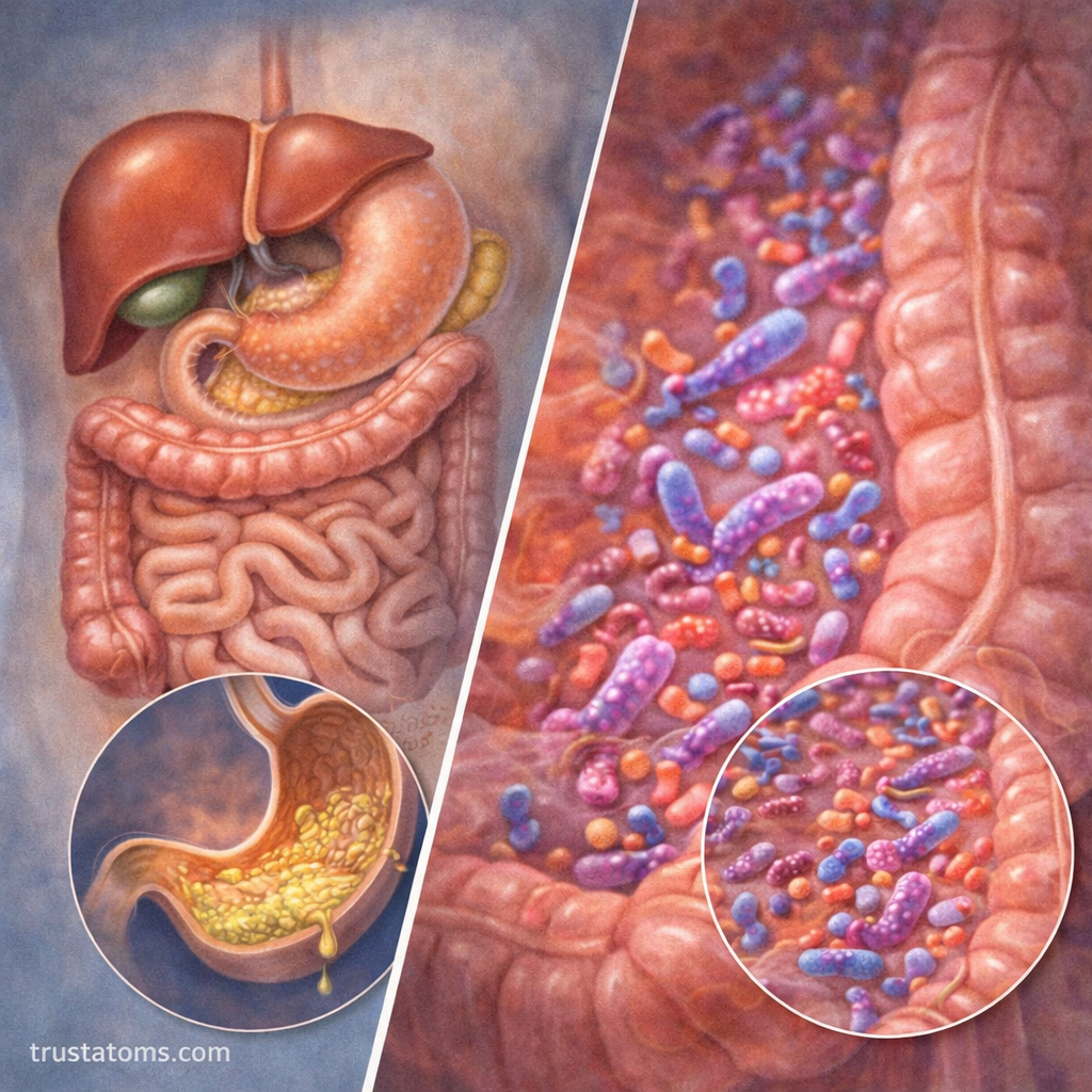
The large intestine receives material that remains after small-intestinal digestion and absorption. It includes the cecum and the ascending, transverse, descending, and sigmoid sections of the colon.
Its lining absorbs remaining water and electrolytes, gradually converting intestinal contents into more solid feces. The process is examined further in Large Intestine Function and Water Absorption.
The colon also contains a dense community of microorganisms. Some ferment undigested dietary fibers, producing short-chain fatty acids that can be absorbed and used by intestinal cells. Certain gut bacteria also synthesize vitamin K and some B vitamins, although the amount available to the body varies.
Rectum and Anus: The Final Stage of Digestion
The rectum temporarily stores feces until elimination. Stretching of the rectal wall activates reflexes associated with the urge to defecate.
The internal anal sphincter operates involuntarily, while the external anal sphincter is under voluntary control. Together with pelvic floor muscles, these structures help regulate the release of feces through the anus.
How Food Moves Through the Digestive Tract
Digestive movement depends on coordinated contractions of smooth muscle. Peristalsis propels contents forward, while segmentation contractions in the intestines mix contents with digestive secretions and expose nutrients to the absorptive lining.
For example, after eating a piece of bread, chewing and saliva begin starch digestion. The swallowed food reaches the stomach, where it is mixed into chyme. In the small intestine, pancreatic enzymes continue starch breakdown, and the resulting glucose is absorbed into the bloodstream. Undigested components continue toward the colon.
Transit time varies with the type of food, meal size, medications, and individual physiology. The entire process can extend over many hours or longer.
The Four Layers of the Digestive Tract Wall
Most of the gastrointestinal tract has four basic tissue layers, although their structures differ by region.
- Mucosa: The innermost layer, involved in protection, secretion, and absorption.
- Submucosa: Contains connective tissue, blood vessels, lymphatic vessels, and nerves.
- Muscularis externa: Produces contractions that mix and propel digestive contents.
- Serosa or adventitia: Forms the outer covering, depending on the organ’s location and surrounding attachments.
These layers explain how the same continuous tract can transport food, secrete digestive substances, absorb nutrients, and protect surrounding tissues.
Common Conditions Affecting Different Digestive Organs
Digestive disorders often affect particular regions. Gastroesophageal reflux involves backward movement of stomach contents into the esophagus. Gastritis affects the stomach lining, while conditions such as celiac disease can damage the small intestine and interfere with absorption.
Constipation, inflammatory bowel disease, and other conditions can affect the colon or rectum. Symptoms sometimes overlap between organs, so their location alone does not establish a diagnosis.
Frequently Asked Questions
What is the correct order of the digestive tract?
The pathway is mouth, pharynx, esophagus, stomach, small intestine, large intestine, rectum, and anus. The liver, gallbladder, and pancreas assist digestion but are not part of the passage food travels through.
Where are most nutrients absorbed?
Most nutrients are absorbed in the small intestine, especially through the specialized surfaces of the jejunum and ileum. The large intestine mainly absorbs remaining water, electrolytes, and certain products of bacterial fermentation.
What is the difference between the small and large intestines?
The small intestine is longer and narrower, with structures specialized for digestion and nutrient absorption. The large intestine is shorter and wider, concentrating intestinal waste through water absorption and supporting microbial fermentation.
The digestive tract combines specialized organs into a continuous pathway, beginning with chewing and swallowing and ending with waste elimination. Its muscular movements, digestive secretions, and absorptive surfaces allow nutrients to move from food into the body while undigested material continues toward the colon and rectum. Understanding these anatomical differences helps explain both normal digestion and the effects of disorders affecting specific regions.
This article is for general education and is not medical advice. Talk to a qualified health professional about any medical concern.
Sources: Gray’s Anatomy for Students; Guyton and Hall Textbook of Medical Physiology; National Institute of Diabetes and Digestive and Kidney Diseases (NIDDK).