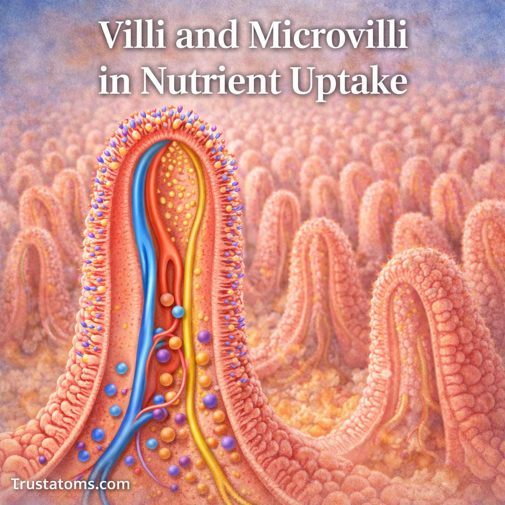
Villi and microvilli increase the surface area of the small intestine, allowing digested nutrients to be absorbed efficiently into the body. Villi are tiny finger-like projections containing blood and lymphatic vessels, while microvilli are even smaller extensions of intestinal cells that support the final stages of digestion and nutrient uptake.
At the center of this process are villi and microvilli, tiny structural adaptations in the small intestine that dramatically increase the surface area available for absorption. Understanding these structures connects microscopic human anatomy with the everyday process of obtaining energy and nutrients from food.
What Are Intestinal Villi?
Villi are microscopic, finger-like projections extending from the inner lining of the small intestine into its hollow center, called the lumen. Each villus increases contact between digested food and the intestinal surface.
A villus is covered by a single layer of epithelial cells, including nutrient-absorbing enterocytes and mucus-producing goblet cells. Beneath this lining are connective tissue, blood capillaries, and a central lymphatic vessel called a lacteal.
These structures connect nutrient absorption to circulation. Nutrient Absorption Pathways into Blood explains how absorbed substances travel from intestinal cells into the body’s transport systems.
The relationship between digestion and blood flow is explored further in Digestive System Integration with Circulation. For the broader route food follows before reaching these structures, see Digestive Tract Anatomy.
What Are Microvilli?
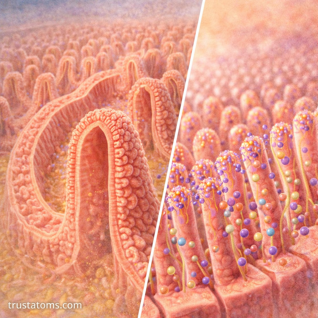
Microvilli are extremely small, densely packed projections extending from the apical surface of enterocytes, the cells responsible for absorbing most nutrients. Unlike villi, which contain several tissue components, microvilli are extensions of individual cells.
Each microvillus contains a supporting framework of actin filaments. Together, thousands of microvilli create a brush-like surface called the brush border.
The brush border provides additional membrane area for nutrient transporters and digestive enzymes. It helps complete the breakdown of certain carbohydrates and proteins before their products enter intestinal cells.
Villi Versus Microvilli: Structural Differences
| Feature | Villi | Microvilli |
|---|---|---|
| Structure | Finger-like projections of intestinal mucosa | Tiny extensions of individual epithelial cells |
| Typical size | Approximately 0.5–1.5 mm long | Approximately 1 micrometer long |
| Location | Inner surface of the small intestine | Apical surface of enterocytes |
| Main function | Increase absorptive surface and support nutrient transport | Expand cell membrane area and support final digestion |
| Blood vessels | Contain capillaries and lacteals | Do not contain blood or lymphatic vessels |
Although both increase surface area, they operate at different structural levels. Villi organize absorption across the intestinal lining, while microvilli expand the working surface of each absorptive cell.
How the Small Intestine Maximizes Surface Area
The small intestine increases its absorptive area through three overlapping structural adaptations:
- Circular folds: Large folds of the intestinal wall increase surface area and influence the movement of intestinal contents.
- Villi: Microscopic projections extend from the mucosal surface, exposing more tissue to digested nutrients.
- Microvilli: Cellular projections create a densely packed brush border with enzymes and transport proteins.
These features provide much more absorptive surface than a smooth intestinal tube of the same length. The familiar comparison to a tennis court is an older estimate; modern measurements generally suggest a smaller total surface area, although estimates vary with the methods used.
How Nutrients Move Through Villi
After food is broken down into absorbable components, nutrients move from the intestinal lumen through epithelial cells and into blood or lymphatic vessels.
- Digestive enzymes break carbohydrates, proteins, and fats into smaller components.
- These products reach the brush border, where certain enzymes complete digestion.
- Nutrients cross the epithelial membrane through specific transport mechanisms.
- Absorbed substances enter capillaries or lymphatic vessels, depending on their properties.
For example, after eating bread, starch is broken down into smaller sugars and eventually glucose. Glucose enters enterocytes through specialized transport proteins, moves into nearby capillaries, and travels through the hepatic portal circulation toward the liver.
Different Nutrients Follow Different Pathways
Carbohydrates and Proteins
Most digestible carbohydrates are absorbed as monosaccharides, including glucose, galactose, and fructose. Protein digestion produces amino acids and small peptides, which enter intestinal cells through specialized transporters.
Most absorbed amino acids subsequently enter the blood. Protein Digestion and Amino Acid Uptake describes the enzymes and transport processes involved.
Dietary Fats
Most long-chain dietary fats are digested into fatty acids and monoglycerides. After absorption, intestinal cells reassemble them into triglycerides and package them into particles called chylomicrons.
Chylomicrons enter lacteals and travel through the lymphatic system before reaching the bloodstream. Shorter-chain fatty acids can enter portal blood more directly.
Vitamins and Minerals
Water-soluble vitamins generally enter blood circulation, while fat-soluble vitamins are absorbed alongside dietary fats. Some nutrients require specialized mechanisms: vitamin B12, for example, is absorbed primarily in the terminal ileum with the help of intrinsic factor.
How the Brush Border Completes Digestion
Many digestive reactions occur before nutrients reach the intestinal lining, but some final steps take place directly at the brush border.
Enzymes such as lactase, sucrase-isomaltase, and maltase-glucoamylase help break down carbohydrates into absorbable sugars. Brush-border peptidases also help process small peptides.
Transport proteins located in the same membrane then move digestion products into enterocytes. This close arrangement allows final digestion and absorption to occur at the same surface.
What Happens When Villi Are Damaged?
Damage to intestinal villi can reduce the surface available for nutrient absorption. When absorption becomes inadequate, the resulting condition is called malabsorption.
- Celiac disease: An immune response to gluten damages the small intestinal lining and can cause villous atrophy, or flattening of the villi.
- Intestinal infections: Certain infections damage epithelial cells or disrupt their absorptive functions.
- Inflammatory disorders: Conditions affecting the small intestine, including some forms of Crohn’s disease, can interfere with nutrient uptake.
Possible consequences include diarrhea, weight loss, bloating, and deficiencies in vitamins or minerals. The effects depend on the affected intestinal region, the extent of damage, and the nutrients involved.
Frequently Asked Questions
Are villi and microvilli the same thing?
No. Villi are projections of intestinal tissue containing blood and lymphatic vessels. Microvilli are much smaller extensions of the epithelial cells covering those villi. Both increase the surface area available for absorption.
Do villi absorb nutrients directly into the blood?
Many nutrients, including glucose and amino acids, pass through intestinal epithelial cells and enter capillaries inside the villi. Most long-chain dietary fats instead enter lymphatic vessels as chylomicrons before reaching the bloodstream.
Can damaged intestinal villi recover?
In some conditions, yes. For example, villi damaged by celiac disease can recover after strict, sustained removal of gluten from the diet. Recovery varies with age, severity, and other factors, and some intestinal conditions cause more persistent damage.
Villi and microvilli make the small intestine an effective absorptive surface by combining extensive membrane area with specialized digestive enzymes and nutrient transporters. Their organization also explains why damage to the intestinal lining can interfere with nutrition: successful absorption depends on healthy epithelial cells and functioning blood and lymphatic pathways.
This article is for general education and is not medical advice. Talk to a qualified health professional about any medical concern.
Sources: Guyton and Hall Textbook of Medical Physiology; Junqueira’s Basic Histology: Text and Atlas; National Institute of Diabetes and Digestive and Kidney Diseases.