
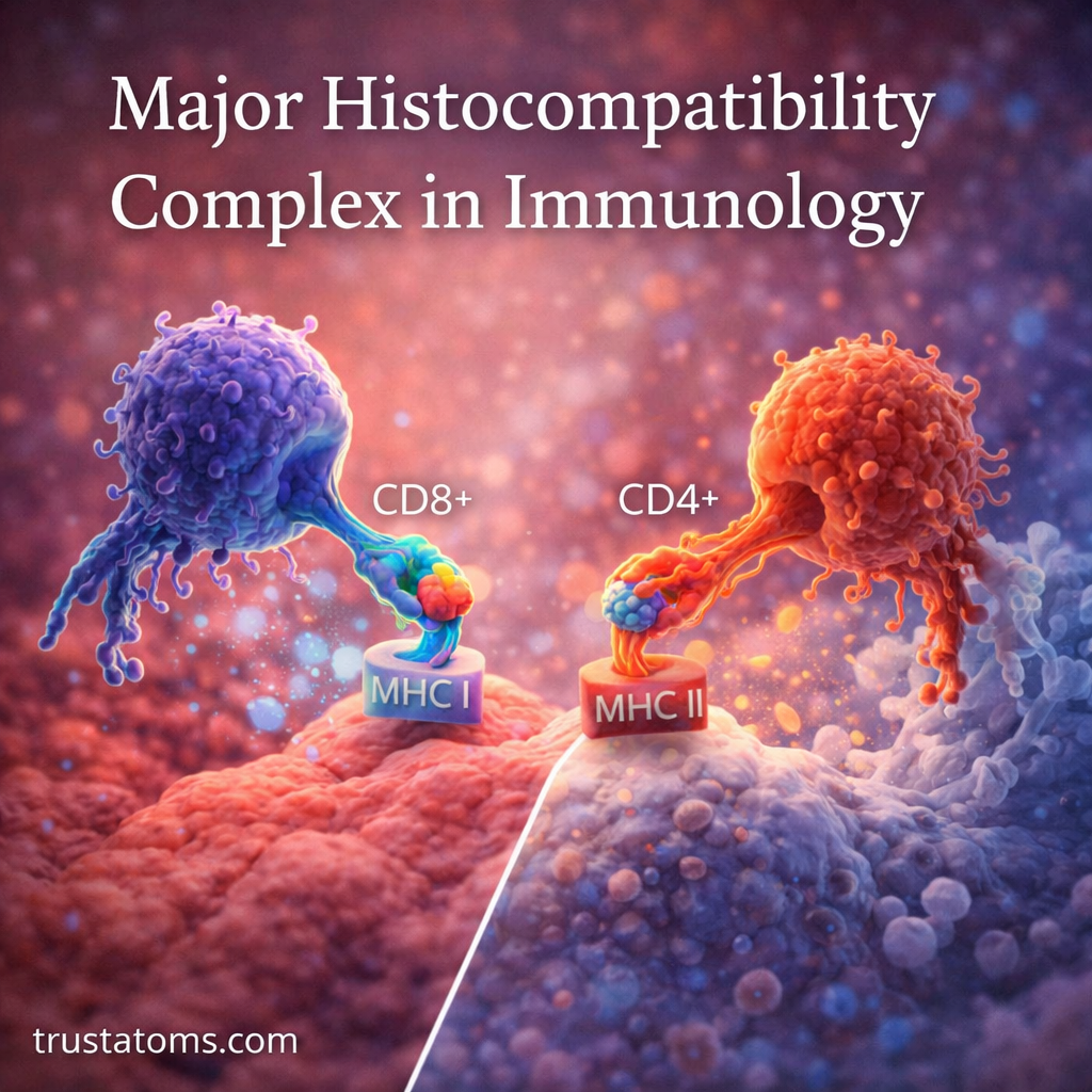
The major histocompatibility complex (MHC) is a group of genes that encode proteins responsible for displaying peptide fragments to T cells, allowing the immune system to examine what is happening inside cells and in surrounding tissues. In humans, these proteins are called human leukocyte antigens (HLA), and they help direct immune responses against infections, abnormal cells, and transplanted tissues.
MHC molecules connect cellular activity with immune recognition, making them an important subject in human anatomy and immunology. Their two principal classes, MHC I and MHC II, display peptides from different sources and interact with different types of T cells.
What Is the Major Histocompatibility Complex?
The major histocompatibility complex is a region of DNA on human chromosome 6 containing genes involved in immune recognition. Among its products are MHC class I and class II proteins, which display short protein fragments, called peptides, on cell surfaces. These peptides may come from the body’s own proteins or from foreign antigens, such as viral proteins.
MHC proteins do not identify danger independently. Instead, they hold peptides in a form that T-cell receptors can recognize. Whether recognition leads to an immune response depends on the T cell, the presented peptide, and additional signals.
Two major functions are displaying peptides from inside cells and presenting material collected from outside cells. These functions allow the immune system to monitor different sources of potential threats.
MHC Class I and Class II: The Main Differences
Although both classes present peptides to T cells, they differ in their distribution, antigen-processing pathways, and the immune cells that recognize them.
| Feature | MHC Class I | MHC Class II |
|---|---|---|
| Typical location | Nearly all nucleated cells | Professional antigen-presenting cells |
| Peptide source | Primarily proteins produced inside cells | Primarily proteins taken up from outside cells |
| T-cell interaction | CD8 T cells | CD4 T cells |
| Main function | Supports recognition of infected or abnormal cells | Supports helper T-cell responses and immune coordination |
| Major human molecules | HLA-A, HLA-B, HLA-C | HLA-DP, HLA-DQ, HLA-DR |
MHC Class I: Monitoring Proteins Inside Cells
MHC class I molecules are expressed on nearly all nucleated cells. They generally present peptides derived from proteins within the cell, including normal cellular proteins and proteins produced during viral infection.
These peptide-MHC complexes can be recognized by CD8 cytotoxic T cells. When an appropriately activated cytotoxic T cell recognizes its target peptide, it may kill the infected or abnormal cell. This is one way MHC molecules help T cells recognize infected or abnormal cells.
MHC Class II: Presenting Material Collected From Outside
MHC class II molecules are mainly expressed by professional antigen-presenting cells, including dendritic cells, macrophages, and B lymphocytes. These cells take up material from their surroundings, break proteins into peptides, and display selected fragments using MHC II.
CD4 helper T cells recognize peptides presented by MHC II. Activated helper T cells can then assist B cells with antibody production, activate macrophages, and coordinate other immune responses.
How MHC Antigen Presentation Works
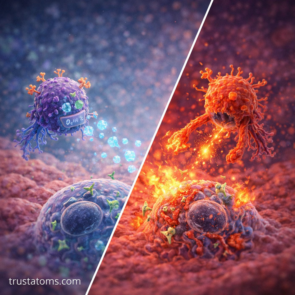
MHC molecules are central to antigen presentation—the process of showing immune cells what to respond to. The two classes use different pathways to prepare peptides for display.
The MHC Class I Processing Pathway
- Proteins inside a cell are broken into peptides, commonly by proteasomes.
- Peptides enter the endoplasmic reticulum through the TAP transporter.
- Suitable peptides bind to newly assembled MHC class I molecules.
- The peptide-MHC complexes travel to the cell surface for examination by CD8 T cells.
The MHC Class II Processing Pathway
- An antigen-presenting cell takes up extracellular material through endocytosis or phagocytosis.
- Proteins are broken into peptides within intracellular compartments.
- MHC class II molecules acquire suitable peptides after removal of the invariant-chain-derived CLIP fragment.
- Peptide-MHC II complexes reach the surface, where CD4 T cells can recognize them.
These are the principal pathways, although specialized processes such as cross-presentation allow some dendritic cells to present externally acquired antigens through MHC class I.
An Example: How MHC Helps Detect a Viral Infection
Imagine a respiratory epithelial cell infected by a virus. As the virus reproduces, the cell produces viral proteins alongside its normal proteins. Some viral proteins are broken into peptides and displayed on MHC class I molecules.
A previously activated CD8 T cell with a matching receptor may recognize one of these viral peptide-MHC complexes and trigger the infected cell’s destruction. Meanwhile, dendritic cells can present viral antigens to helper T cells, supporting broader immune coordination.
This example demonstrates why MHC molecules are more accurately described as peptide-display platforms than simple cellular identification tags. They provide information that T cells interpret through highly specific receptors.
MHC and Self-Recognition
MHC molecules routinely display peptides derived from the body’s own proteins. Healthy immune function requires T cells to recognize MHC while generally avoiding harmful responses against normal self-peptides.
During T-cell development in the thymus, positive selection helps retain cells capable of recognizing self-MHC molecules, while negative selection eliminates many cells that react too strongly to self-antigens.
Additional tolerance mechanisms operate outside the thymus. These safeguards are discussed in Immune Tolerance and Self-Recognition.
Self-tolerance is not absolute. Failures in these mechanisms can contribute to autoimmune disease, in which immune responses damage the body’s own tissues.
Why MHC Genes Vary Between Individuals
Human HLA genes are highly polymorphic, meaning many different versions, or alleles, exist within the population. Individuals inherit HLA variants from both parents and generally express molecules from both inherited sets.
Different HLA molecules bind different collections of peptides. This variation affects which antigens can be presented effectively and contributes to differences in immune responses between individuals.
HLA diversity also means that two unrelated people may have substantially different tissue compatibility profiles, an important consideration in transplantation.
MHC and Organ Transplantation
HLA differences between donors and recipients can contribute to transplant rejection because recipient immune cells may recognize donor HLA molecules or associated peptides as foreign.
HLA typing helps assess compatibility, especially for hematopoietic stem cell transplantation and certain solid-organ transplants. Matching requirements vary by transplant type, and clinicians also consider blood-group compatibility, antibodies, and other medical factors.
Even with favorable matching, immunosuppressive treatment is often necessary to reduce rejection risk. HLA compatibility improves risk assessment but cannot guarantee transplant success.
MHC and Disease Susceptibility
Some HLA variants are associated with increased susceptibility to particular immune-mediated conditions. For example, HLA-B27 is strongly associated with ankylosing spondylitis, while certain HLA-DQ variants are associated with celiac disease and type 1 diabetes.
These associations do not mean that everyone carrying a particular HLA variant will develop the associated condition. Genetic background, environmental exposures, and other immune mechanisms also influence disease risk.
MHC and the Adaptive Immune System
MHC-dependent peptide recognition is central to many adaptive immune responses. However, recognizing a peptide-MHC complex is not always sufficient to activate a naive T cell. Additional costimulatory signals and cytokines help determine whether activation, tolerance, or another outcome occurs.
These additional requirements are explained in Adaptive Response Activation Pathways.
Once appropriately activated, T cells can multiply, perform specialized functions, and generate memory populations. MHC molecules therefore connect antigen recognition with the broader processes of adaptive immune defense.
Frequently Asked Questions
What is the difference between MHC and HLA?
MHC is the general term for the genetic region and associated antigen-presenting molecules. HLA, or human leukocyte antigen, is the name used for the corresponding system in humans.
Do all human cells have MHC molecules?
Nearly all nucleated human cells express MHC class I molecules. Mature red blood cells lack nuclei and normally do not express MHC class I. MHC class II expression is mainly associated with professional antigen-presenting cells, although it can be induced in some other cells under particular conditions.
Can MHC molecules directly kill infected cells?
No. MHC molecules display peptides but do not directly destroy infected cells. Appropriately activated cytotoxic T cells can recognize specific peptide-MHC class I complexes and initiate mechanisms that kill their target cells.
The major histocompatibility complex provides the molecular framework through which T cells examine peptides displayed by other cells. MHC class I primarily supports CD8 T-cell recognition, while MHC class II supports CD4 T-cell responses. Differences in HLA genes also help explain variations in immune recognition, disease associations, and transplant compatibility.
This article is for general education and is not medical advice. Talk to a qualified health professional about any medical concern.
Sources: Abbas, Lichtman, and Pillai, Cellular and Molecular Immunology; Janeway’s Immunobiology; National Cancer Institute.