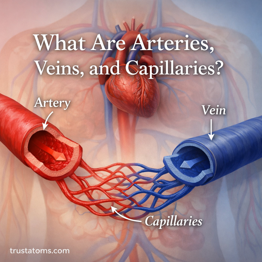
Arteries are blood vessels that carry blood away from the heart, veins return blood toward the heart, and capillaries are tiny vessels where oxygen, nutrients, and waste products move between blood and surrounding tissues. Their different wall structures and functions allow blood to travel efficiently through the body while supporting the needs of individual cells.
These three vessel types form a connected network rather than separate systems. Understanding their differences is a useful part of studying human anatomy because blood vessels connect the heart, lungs, brain, muscles, and nearly every other living tissue.
A simple way to remember their functions is that arteries distribute blood, capillaries provide exchange surfaces, and veins collect returning blood. However, whether a vessel carries oxygen-rich or oxygen-poor blood depends on where it is located in the circulation.
Overview of Blood Vessels in the Circulatory System
The human circulatory system is a vast and efficient network responsible for transporting blood, oxygen, nutrients, and waste products throughout the body. The heart provides the pumping force, while blood vessels distribute blood through connected pathways.
Blood moves through two main circuits: pulmonary circulation between the heart and lungs, and systemic circulation between the heart and body tissues. Their routes and functions are compared in Systemic vs Pulmonary Circulation.
Pressure and resistance influence how blood moves through different vessels. Larger arteries carry pulsating blood flow from the heart, while smaller vessels create much of the resistance that helps regulate tissue perfusion. These principles are explored in Hemodynamics in Large Vessels.
Some vessels serve particular organs. For example, coronary arteries supply oxygenated blood to the heart muscle itself, as explained in Coronary Circulation and Heart Supply.
At the smallest scale, blood flow through arterioles, capillaries, and venules supports exchange with tissues. This local network is called microcirculation in tissue systems.
What Are Arteries?
Arteries are vessels that carry blood away from the heart. In systemic circulation, they generally transport oxygen-rich blood from the left ventricle toward body tissues. In pulmonary circulation, arteries carry oxygen-poor blood from the right ventricle toward the lungs.
Arteries usually have relatively thick walls containing smooth muscle and elastic connective tissue. These features help them withstand pressure and regulate blood flow.
How Arterial Walls Handle Pressure
The walls of most arteries contain three main layers: an inner tunica intima, a middle tunica media, and an outer tunica externa. The tunica media contains smooth muscle and varying amounts of elastic tissue.
Large elastic arteries, including the aorta, expand when the heart ejects blood and recoil between heartbeats. This helps smooth the pulsating output of the heart. Smaller muscular arteries adjust their diameter to influence blood distribution.
Examples of Major Arteries
- Aorta: The largest artery, carrying blood from the left ventricle into systemic circulation.
- Carotid arteries: Supply blood to structures in the head and neck, including the brain through the internal carotid arteries.
- Coronary arteries: Deliver oxygenated blood to the heart muscle.
- Pulmonary arteries: Carry oxygen-poor blood from the heart toward the lungs.
As arteries branch, they become progressively smaller. Arterioles, the small vessels leading toward capillary networks, can change their diameter through smooth muscle contraction and relaxation, helping control local blood flow.
What Are Veins?
Veins carry blood toward the heart. Most systemic veins transport oxygen-poor blood from body tissues, while pulmonary veins return oxygen-rich blood from the lungs to the left atrium.
Compared with arteries of similar size, veins generally have thinner walls and larger internal spaces, called lumens. Their walls contain less smooth muscle and elastic tissue because venous blood usually flows under much lower pressure.
How Veins Return Blood at Low Pressure
Venous return depends on pressure differences, the pumping action of the heart, and several supporting mechanisms. When skeletal muscles contract, they compress nearby veins and help move blood toward the heart.
Many veins in the limbs contain one-way valves that reduce backward flow. These valves are especially useful in the legs, where blood must return upward against gravity while a person is standing.
Not all veins have valves. Large central veins and some other venous pathways lack them, so the presence of valves is a common feature rather than a universal definition.
Superficial, Deep, and Pulmonary Veins
- Superficial veins: Located closer to the skin and often visible on the hands or arms.
- Deep veins: Located deeper within the body, frequently alongside arteries and between muscles.
- Pulmonary veins: Carry oxygen-rich blood from the lungs to the left atrium.
- Venae cavae: Large veins that return systemic blood to the right atrium.
Veins also act as blood reservoirs because their compliant walls allow them to hold substantial volumes of blood at relatively low pressure.
What Are Capillaries?
Capillaries are microscopic blood vessels that connect the arterial and venous sides of local circulation. Their walls consist mainly of a single layer of endothelial cells supported by a basement membrane, creating a short distance for exchange.
Many capillaries are approximately 5–10 micrometers in diameter, although dimensions vary among tissues. Their narrow lumens often require red blood cells to deform as they pass through.
How Capillaries Exchange Substances
Oxygen and carbon dioxide move mainly by diffusion, following differences in their partial pressures. Nutrients and other small substances can also cross capillary walls through mechanisms that depend on the substance and tissue.
Fluid movement is influenced by pressure differences and the properties of the capillary barrier. The lymphatic system returns excess interstitial fluid to the circulation, helping maintain tissue fluid balance.
Three Types of Capillaries
- Continuous capillaries: Have a relatively uninterrupted endothelial lining and occur in tissues such as skeletal muscle and the central nervous system.
- Fenestrated capillaries: Contain small openings that support exchange in organs such as the kidneys and intestinal lining.
- Sinusoidal capillaries: Have larger openings and more permeable walls, allowing certain large substances or cells to pass in organs such as the liver and bone marrow.
Capillary structure is therefore adapted to the exchange requirements of individual organs rather than being identical throughout the body.
Arteries vs Veins vs Capillaries: Key Differences
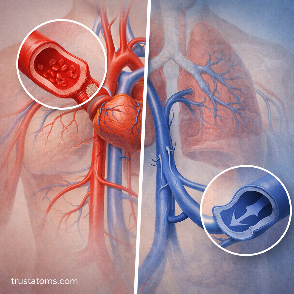
Arteries, veins, and capillaries differ most clearly in blood flow direction, wall thickness, pressure, and their contribution to circulation.
| Feature | Arteries | Veins | Capillaries |
|---|---|---|---|
| Main function | Carry blood away from the heart | Return blood toward the heart | Exchange substances with tissues |
| Blood flow direction | Away from heart | Toward heart | From arterioles toward venules |
| Wall structure | Relatively thick, muscular, elastic | Relatively thin, compliant | Primarily one endothelial cell layer |
| Typical pressure | Higher | Lower | Lower than arterial pressure |
| Valves | Generally absent within arteries | Present in many limb veins | Absent |
| Typical oxygen content in systemic circulation | Oxygen-rich | Oxygen-poor | Changes during exchange |
| Special feature | Withstand and regulate pressure | Store blood and support venous return | Provide short exchange distances |
These comparisons describe general patterns. Vessel structure varies with location and size, and pulmonary circulation reverses the usual oxygen-content pattern of systemic arteries and veins.
How Blood Travels Through All Three Vessel Types
Arteries, veins, and capillaries operate as parts of one continuous circulation. A complete journey includes the heart, lungs, and body tissues.
- The left ventricle pumps oxygen-rich blood into the aorta.
- Systemic arteries branch into smaller arteries and arterioles.
- Blood enters tissue capillaries, where oxygen and nutrients can move toward surrounding cells.
- Blood collects in venules and then larger systemic veins.
- The superior and inferior venae cavae return blood to the right atrium.
- The right ventricle sends blood through pulmonary arteries to lung capillaries for gas exchange.
- Pulmonary veins return oxygen-rich blood to the left atrium, completing the route back toward the left ventricle.
Blood continuously moves through these connected pathways, but individual tissues receive different amounts of blood according to their metabolic needs and the regulation of local vessels.
Worked Example: Why Capillaries Slow Blood Flow
Blood flow becomes much slower in capillary networks than in the aorta, even though the same overall volume of blood must pass through the systemic circuit over time. The explanation is the combined cross-sectional area of the vessels.
Consider a simplified example in which blood moves through a large vessel with a total cross-sectional area of 5 square centimeters at a velocity of 20 centimeters per second.
- Multiply area by velocity: 5 square centimeters × 20 centimeters per second = 100 cubic centimeters per second.
- Imagine the same volume flowing through a capillary network with a combined cross-sectional area of 2,000 square centimeters.
- Divide flow by area: 100 ÷ 2,000 = 0.05 centimeters per second.
- The average velocity is therefore 400 times lower in the simplified capillary network than in the original vessel.
These are illustrative values, not measurements of a particular person. The calculation demonstrates the relationship between flow, total cross-sectional area, and average velocity. Slow capillary flow supports exchange by increasing the time blood spends near surrounding tissues.
Why Blood Vessel Structure Affects Health
Changes in vessel structure or function can interfere with normal circulation. The consequences depend on whether arteries, veins, or capillaries are affected.
- Atherosclerosis: Plaque accumulation in arterial walls can narrow vessels and reduce blood supply to organs.
- Hypertension: Persistently elevated arterial pressure can damage blood vessels and increase cardiovascular risk.
- Varicose veins: Enlarged, twisted superficial veins can develop when venous valves and walls do not maintain efficient blood return.
- Deep vein thrombosis: A blood clot in a deep vein can obstruct flow and may cause serious complications if part of the clot travels to the lungs.
- Microvascular damage: Disorders affecting small vessels can impair tissue exchange, including in the kidneys, eyes, and nerves.
Regular physical activity, avoiding tobacco, managing blood pressure, and following appropriate medical guidance can support vascular health. These measures reduce certain risks but cannot prevent every blood vessel disorder.
Common Misconceptions About Blood Vessels
Misconception 1: Arteries Always Carry Oxygen-Rich Blood
Arteries are defined by carrying blood away from the heart. Pulmonary arteries normally carry oxygen-poor blood toward the lungs, while pulmonary veins return oxygen-rich blood to the heart.
Misconception 2: All Veins Have Valves
Many veins in the arms and legs contain valves, but some major veins do not. Venous return also depends on pressure differences, muscle contractions, and changes in pressure during breathing.
Misconception 3: Blood Flows Directly From Arteries Into Veins
In most tissues, arteries branch into arterioles, which supply capillary networks. Blood then enters venules before returning through larger veins. Some specialized vascular connections can bypass typical capillary pathways.
Misconception 4: Veins Are Blue Because Their Blood Is Blue
Human blood is red, although oxygen-poor blood is generally darker than oxygen-rich blood. Veins can appear blue through the skin because of how light is absorbed and scattered by skin and underlying tissues.
Frequently Asked Questions
What Is the Main Difference Between Arteries, Veins, and Capillaries?
Arteries carry blood away from the heart, veins return blood toward the heart, and capillaries provide thin exchange surfaces between blood and tissues. Their walls differ in thickness and composition according to these functions.
Why Are Artery Walls Thicker Than Vein Walls?
Arteries generally experience higher pressures and contain more smooth muscle and elastic tissue. Veins operate under lower pressure and have more compliant walls that allow them to hold substantial blood volumes.
Do Capillaries Carry Oxygenated or Deoxygenated Blood?
Capillaries can contain blood with different oxygen levels depending on their location and the exchange occurring. Systemic capillary blood generally loses oxygen to tissues, while pulmonary capillary blood gains oxygen from the lungs.
Why Do Veins Have Valves?
Valves in many peripheral veins help prevent backward blood flow, particularly when blood must travel upward against gravity. They work with skeletal muscle contractions to support venous return.
Which Blood Vessels Have the Lowest Blood Pressure?
Pressure generally decreases as blood moves through systemic arteries, arterioles, capillaries, and veins. Large veins near the right atrium normally have some of the lowest pressures in the systemic circulation.
Arteries distribute blood under pressure, capillaries provide the thin surfaces needed for exchange, and veins return blood toward the heart at lower pressure. Their different structures make it possible to transport blood efficiently while meeting the needs of individual tissues. Understanding how these vessels connect also explains why problems in one part of the vascular network can affect circulation throughout the body.
This article is for general education and is not medical advice. Talk to a qualified health professional about any medical concern.
Sources: OpenStax, Anatomy and Physiology 2e; Guyton and Hall, Textbook of Medical Physiology; Tortora and Derrickson, Principles of Anatomy and Physiology; National Heart, Lung, and Blood Institute (NHLBI), How the Heart Works.