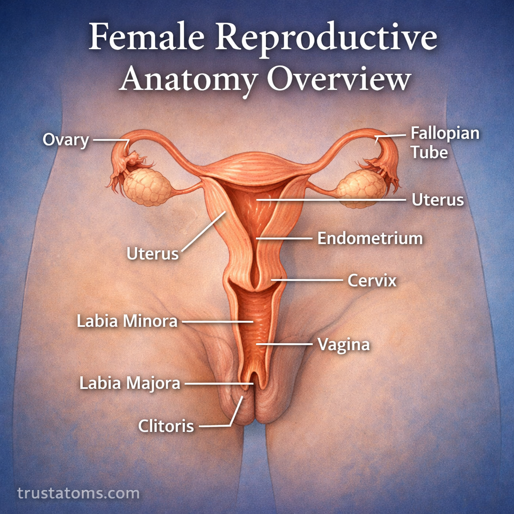
The female reproductive system is a complex and highly coordinated group of organs responsible for producing eggs, supporting fertilization, and nurturing the development of a fetus. Its main structures include the ovaries, fallopian tubes, uterus, cervix, vagina, and vulva, which work together through hormonal signals and specialized anatomical functions.
Understanding this reproductive system connects the study of human anatomy with menstruation, fertility, pregnancy, and reproductive health. Each organ has a distinct structure, but reproductive processes depend on their coordinated activity.
Primary Functions of the Female Reproductive System
The reproductive organs perform several related functions, from producing egg cells to supporting pregnancy and childbirth.
- Egg production (oogenesis): Developing egg cells within ovarian follicles and releasing a mature egg during ovulation.
- Fertilization support: Providing a pathway where sperm can reach an egg, usually within a fallopian tube.
- Pregnancy support: Preparing the uterine lining for implantation and supporting embryo and fetal development.
- Hormone regulation: Producing hormones like estrogen and progesterone that regulate the menstrual cycle and reproductive tissues.
These functions involve both internal reproductive organs and external structures that provide protection, sensation, and access to the reproductive tract.
External Female Reproductive Anatomy: The Vulva
The vulva refers to the external genital structures. It is anatomically distinct from the vagina, which is an internal muscular canal.
The main structures of the vulva include:
- Labia majora: Outer folds of skin that help protect the structures within the vulva.
- Labia minora: Inner folds surrounding the vestibule, where the urethral and vaginal openings are located.
- Clitoris: A highly sensitive erectile organ involved primarily in sexual sensation and arousal.
- Vaginal opening: The external entrance to the vaginal canal.
- Vestibular glands: Glands that contribute secretions to the surrounding tissues.
The external urethral opening is also located in the vestibule, but the urethra belongs to the urinary system rather than the reproductive tract. This distinction helps explain why urination and menstruation use separate anatomical passages.
Internal Female Reproductive Organs
The internal reproductive organs are located mainly within the pelvis. Their positions and tissue structures allow eggs, sperm, and reproductive hormones to participate in different stages of reproduction.
Vagina: A Muscular Canal
The vagina is an elastic, muscular canal extending from the vaginal opening to the cervix. Its walls contain folds that allow stretching, and its lining is adapted to a normally acidic environment during the reproductive years.
The vagina allows menstrual fluid to leave the body, receives semen during vaginal intercourse, and forms part of the birth canal during vaginal delivery.
Cervix: The Lower Portion of the Uterus
The cervix is the narrow, lower portion of the uterus that projects into the upper vagina. Its central passage, the cervical canal, connects the uterine cavity with the vagina.
Cervical mucus changes throughout the menstrual cycle. Around ovulation, it generally becomes thinner and more favorable for sperm movement. During childbirth, the cervix softens and dilates to permit passage of the baby.
Uterus: Where Pregnancy Develops
The uterus is a hollow, muscular organ usually located between the urinary bladder and rectum. It contains three main tissue layers: the endometrium, myometrium, and perimetrium.
- Endometrium: The inner lining that changes during the menstrual cycle and provides the site for implantation.
- Myometrium: The thick smooth-muscle layer responsible for uterine contractions.
- Perimetrium: The outer covering of much of the uterus.
During pregnancy, the uterus enlarges substantially as the fetus develops. During labor, coordinated contractions of the myometrium help move the baby through the cervix and vagina.
Fallopian Tubes: Transport and Fertilization
The fallopian tubes, also called uterine tubes or oviducts, extend from the upper uterus toward the ovaries. Their fingerlike fimbriae help capture the egg after ovulation.
Cilia and smooth-muscle contractions help transport the egg or early embryo toward the uterus. Fertilization most commonly occurs in the ampulla, the wider middle portion of a fallopian tube.
Ovaries: Egg Development and Hormone Production
The ovaries are paired reproductive glands located on either side of the uterus. They contain follicles in different stages of development, each surrounding an immature egg cell.
During reproductive years, ovarian follicles produce estrogen. After ovulation, the remaining follicular tissue forms the corpus luteum, which produces progesterone and estrogen to support the uterine lining.
The Menstrual Cycle: How the Organs Work Together
The menstrual cycle coordinates ovarian activity with changes in the uterine lining. A 28-day cycle is a common teaching example, although adult menstrual cycles often range from approximately 21 to 35 days.
The main stages are:
- Menstruation: The uterine lining sheds, producing menstrual bleeding.
- Follicular phase: Ovarian follicles develop, estrogen generally rises, and the uterine lining rebuilds.
- Ovulation: Release of a mature egg from the ovary, followed by its movement toward the fallopian tube.
- Luteal phase: Progesterone secretion increases as the corpus luteum supports the uterine lining.
Menstruation overlaps with the beginning of the follicular phase rather than occurring as an entirely separate ovarian stage. For a closer look at the timing of these changes, see Menstrual Cycle Phases.
Hormonal Control of Female Reproduction
Reproductive hormones are coordinated through communication between the hypothalamus, pituitary gland, and ovaries. This network is called the hypothalamic-pituitary-ovarian axis.
| Hormone | Main Function |
|---|---|
| FSH | Stimulates ovarian follicle development. |
| LH | Triggers ovulation and supports corpus luteum formation. |
| Estrogen | Promotes uterine lining growth and regulates reproductive tissues. |
| Progesterone | Prepares and maintains the endometrium after ovulation. |
These hormones interact through feedback mechanisms. For example, sustained high estrogen levels near the end of the follicular phase stimulate an LH surge that triggers ovulation.
Fertilization and Early Pregnancy
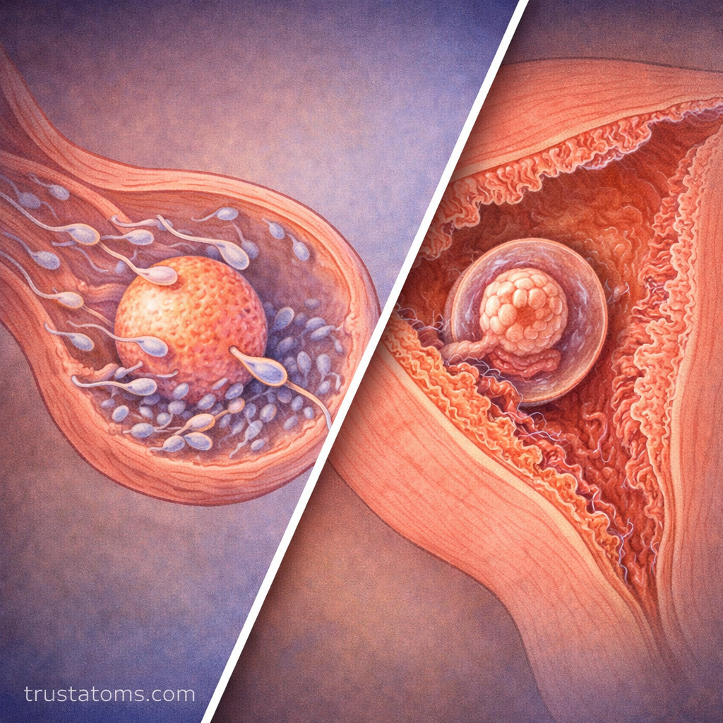
If sperm encounters an egg within a fallopian tube, fertilization may occur. The resulting zygote begins dividing as it travels toward the uterus, developing into a blastocyst before implantation.
As a concrete example, fertilization typically occurs within about 24 hours after ovulation if viable sperm are present. Implantation generally begins approximately 6–7 days after fertilization, although the exact timing varies.
After implantation, developing placental tissue produces human chorionic gonadotropin (hCG), which helps maintain progesterone production by the corpus luteum during early pregnancy.
Common Conditions Affecting Reproductive Anatomy
Several conditions can affect reproductive organs, menstrual function, or fertility:
- Endometriosis: Tissue resembling the uterine lining grows outside the uterus and may cause pelvic pain or fertility difficulties.
- Uterine fibroids: Noncancerous growths of uterine muscle tissue that may contribute to heavy bleeding or pelvic pressure.
- Polycystic ovary syndrome: A hormonal and metabolic condition commonly associated with irregular ovulation.
- Cervical dysplasia: Abnormal cervical cell changes, often associated with persistent infection by certain human papillomavirus types.
- Ectopic pregnancy: Implantation outside the uterine cavity, most commonly within a fallopian tube, requiring prompt medical attention.
Knowledge of normal anatomy helps explain where symptoms originate and why pelvic examinations, cervical screening, or imaging may be recommended in particular situations.
Frequently Asked Questions
What is the difference between the vulva and vagina?
The vulva consists of the external genital structures, including the labia and clitoris. The vagina is the internal muscular canal extending from the vaginal opening to the cervix.
Where does fertilization normally occur?
Fertilization most commonly occurs in the ampulla of a fallopian tube. The early embryo then travels toward the uterus, where implantation may occur in the endometrium.
Do the ovaries produce new eggs every month?
No. The ovaries contain a finite population of immature egg cells established before birth. During each cycle, a group of follicles begins developing, and typically one releases an egg during ovulation.
Female reproductive anatomy combines specialized structures for egg development, hormone secretion, fertilization, pregnancy, and childbirth. The ovaries and fallopian tubes support egg release and transport, while the uterus, cervix, vagina, and vulva contribute distinct functions that change throughout the reproductive cycle.
This article is for general education and is not medical advice. Talk to a qualified health professional about any medical concern.
Sources: Gray’s Anatomy for Students; Williams Gynecology; American College of Obstetricians and Gynecologists (ACOG).