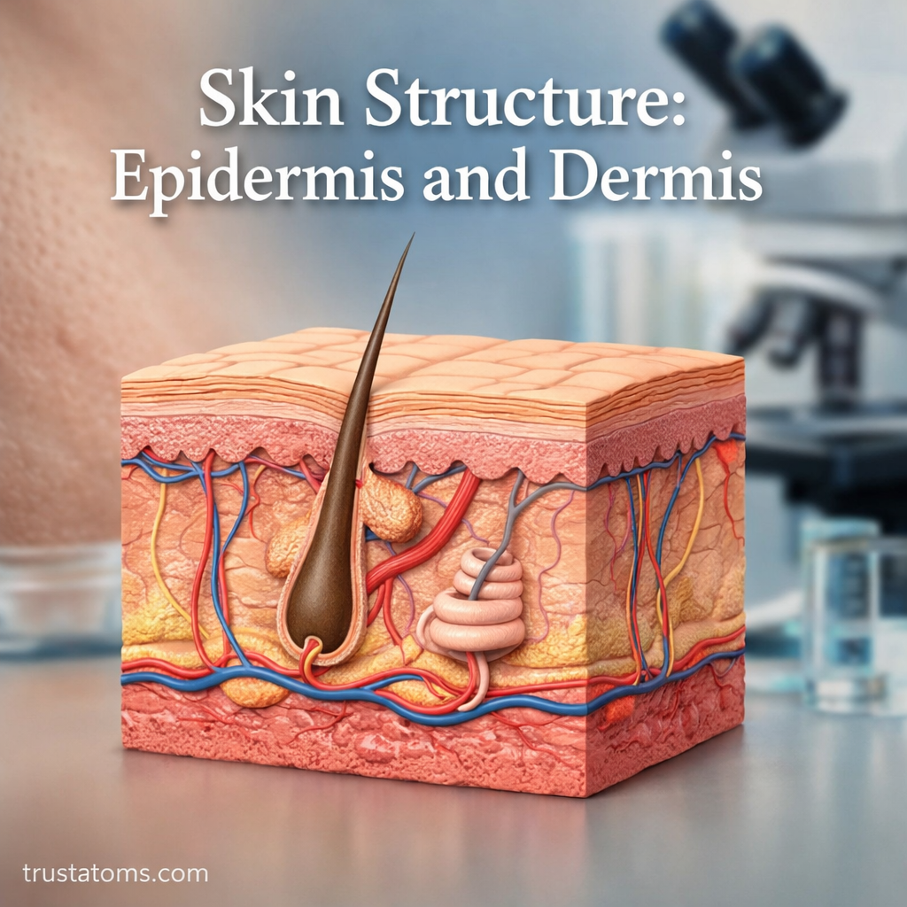
Human skin has two main layers: the epidermis, a thin outer barrier that limits water loss and protects against environmental damage, and the dermis, a deeper layer that provides strength, sensation, and support. Beneath them lies the hypodermis, a layer of subcutaneous tissue that cushions the body and helps insulate it.
Skin is the body’s largest organ and a useful example of how structures in human anatomy are adapted to different functions. Its layers work together to protect underlying tissues, regulate temperature, detect sensations, and repair everyday injuries.
Overview of Skin Layers
The skin and its underlying tissues are commonly described in three layers, arranged from the surface inward:
- Epidermis: The outer epithelial layer, made primarily of keratinocytes that form a protective, water-resistant barrier.
- Dermis: The connective tissue layer containing collagen, elastic fibers, blood vessels, nerves, hair follicles, and glands.
- Hypodermis: The underlying subcutaneous tissue, composed largely of fat and connective tissue. It provides cushioning, insulation, and attachment to deeper structures.
Strictly speaking, the epidermis and dermis form the skin itself, while the hypodermis lies beneath it. The arrangement of cells and lipids in the epidermis is central to skin barrier function and protection.
Structures associated with the dermis perform specialized tasks. Sebaceous glands and skin oils help lubricate the skin and hair, while sweat glands and thermoregulation explain how sweating contributes to cooling the body.
What Is the Epidermis?
The epidermis is the outermost layer of skin. It consists mainly of keratinocytes, which mature as they move toward the surface and eventually form flattened, dead cells that are shed. The epidermis contains no blood vessels; its living cells receive nutrients by diffusion from the underlying dermis.
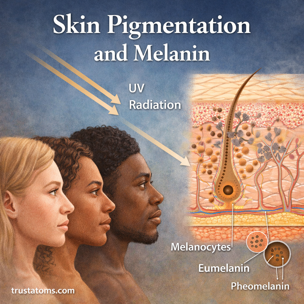
The epidermis also contains melanocytes, which produce the pigment melanin, immune cells called Langerhans cells, and Merkel cells associated with touch sensation. Together with hair, nails, and skin glands, these structures belong to the integumentary system.
The Five Epidermal Layers
In thick skin, such as the palms and soles, the epidermis has five distinct strata, listed from deepest to most superficial:
- Stratum basale: The deepest layer, where dividing cells replenish keratinocytes. Melanocytes are also found here.
- Stratum spinosum: Several layers of keratinocytes connected by strong cell junctions that help resist mechanical stress.
- Stratum granulosum: Cells undergo changes that help establish the epidermis’s protective lipid barrier.
- Stratum lucidum: A thin, translucent layer present in thick skin but generally absent from thin skin.
- Stratum corneum: The outermost layer of flattened, dead, keratin-filled cells embedded in protective lipids.
Most skin has four recognizable epidermal strata because it lacks a distinct stratum lucidum. Epidermal thickness also varies by location, with thicker protective surfaces where friction is greatest.
What Is the Dermis?
The dermis lies directly below the epidermis and is generally much thicker. Its connective tissue contains collagen fibers that resist pulling forces and elastic fibers that help skin return toward its original shape after stretching.
Unlike the epidermis, the dermis contains blood vessels. These vessels supply oxygen and nutrients to dermal tissues, support the epidermis through diffusion, and help control heat loss by changing blood flow near the surface.
The Two Dermal Layers
- Papillary dermis: The superficial region of loose connective tissue. It forms projections called dermal papillae and contains small blood vessels and sensory structures.
- Reticular dermis: The deeper, thicker region containing dense irregular connective tissue, larger vessels, nerves, and many skin appendages.
Hair follicles, sweat glands, sebaceous glands, and sensory nerve endings extend through or occupy portions of the dermis. Some glandular structures and hair follicles can extend into the underlying subcutaneous tissue.
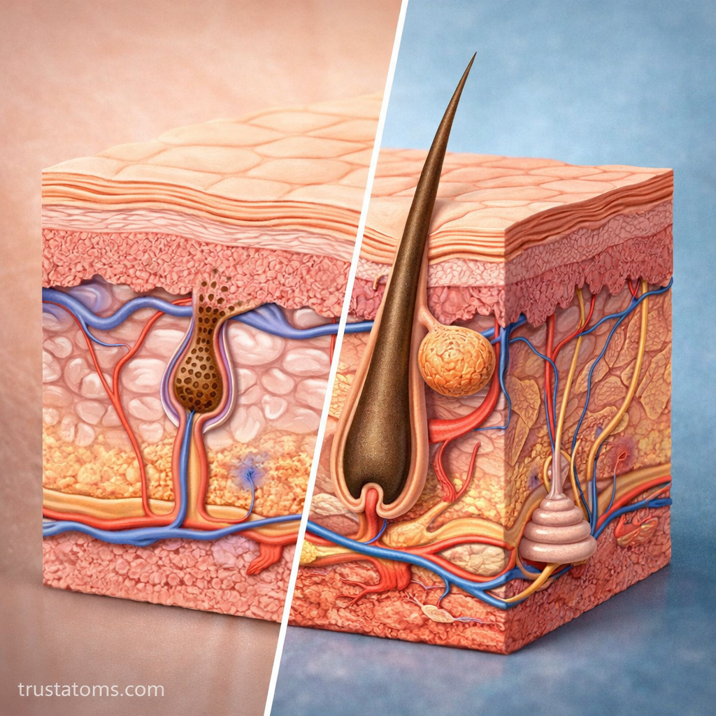
Epidermis vs. Dermis: Main Differences
| Feature | Epidermis | Dermis |
|---|---|---|
| Location | Outer skin layer | Beneath epidermis |
| Main tissue | Keratinized stratified squamous epithelium | Connective tissue |
| Blood vessels | Absent | Present |
| Main function | Barrier protection | Strength, support, and sensation |
| Major components | Keratinocytes, melanocytes, immune cells | Collagen, elastic fibers, vessels, nerves, glands |
| Response to injury | Superficial damage can repair without scarring | Deeper damage may produce scar tissue |
How the Two Layers Work Together
The epidermis and dermis meet at the dermal-epidermal junction, where specialized structures anchor the layers together. The dermis supplies nutrients to living epidermal cells, while the epidermis limits the entry of harmful substances and reduces water loss.
Consider a shallow scrape on your knee. If the injury affects only the epidermis, it may produce little bleeding because that layer has no blood vessels. A deeper cut reaching the dermis can damage capillaries, cause bleeding, and require more extensive tissue repair. Whether a wound scars depends partly on its depth and the extent of dermal damage.
How Skin Structure Affects Everyday Health
The distinction between epidermis and dermis helps explain common skin conditions and normal changes:
- Sunburn: Ultraviolet radiation damages skin cells, particularly in the epidermis, triggering inflammation that can extend into deeper tissue.
- Acne: Blocked and inflamed hair follicles and associated sebaceous glands produce common acne lesions.
- Burns: Superficial burns involve the epidermis, while partial-thickness burns extend into the dermis.
- Skin aging: Changes in dermal collagen and elastic fibers contribute to wrinkles and reduced firmness.
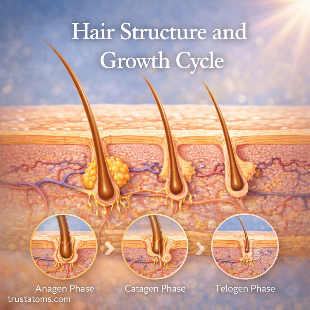
Skin appendages also depend on this layered organization. The hair structure and growth cycle illustrate how follicles embedded in deeper tissue produce hair that emerges through the epidermal surface.
Frequently Asked Questions
Which is thicker, the epidermis or dermis?
The dermis is generally much thicker than the epidermis. Thickness varies with body location, age, and individual characteristics. The epidermis is especially thick on the palms and soles, where the skin experiences repeated friction.
Does the epidermis contain nerves or blood vessels?
The epidermis has no blood vessels, but it does contain sensory nerve endings. Blood vessels remain in the underlying dermis, where they support nearby tissues and contribute to temperature regulation.
Can damaged skin grow back?
Yes. The epidermis continually replaces cells, and superficial injuries can often heal without permanent scarring. Deeper injuries involving substantial dermal damage may heal with scar tissue rather than restoring the original structure completely.
The epidermis provides the skin’s renewable outer barrier, while the dermis supplies the connective tissue, circulation, and sensory structures beneath it. Understanding where these layers differ makes it easier to explain why superficial scrapes, deeper cuts, burns, and changes in skin elasticity affect the body differently.
This article is for general education and is not medical advice. Talk to a qualified health professional about any medical concern.
Sources: OpenStax, Anatomy and Physiology 2e; National Cancer Institute, SEER Training Modules; Merck Manual, Structure and Function of the Skin.