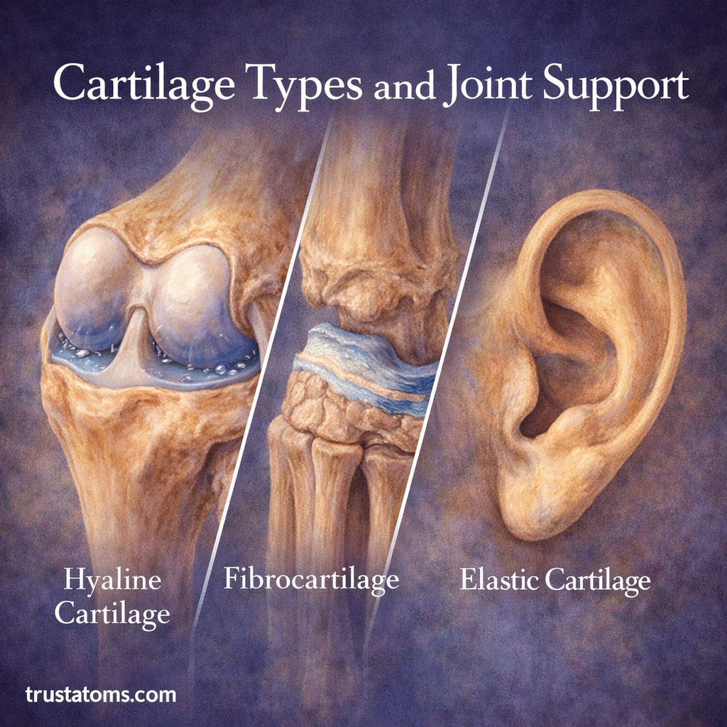
Cartilage Types and Joint Support

Illustration showing hyaline cartilage in a joint, fibrocartilage in the knee, and elastic cartilage in the ear.
Visual comparison of cartilage types and their roles in joint support and flexibility.

Cartilage is a specialized connective tissue that plays a critical role in joint function, flexibility, and shock absorption. Unlike bone, cartilage is smooth, flexible, and designed to reduce friction between moving parts of the body.

There are three main types of cartilage—hyaline, fibrocartilage, and elastic cartilage—each with a different structure and function. Understanding these tissues is part of human anatomy and helps explain how joints withstand pressure, move smoothly, and maintain their shape during everyday activities.

Continue reading “Cartilage Types and Joint Support”
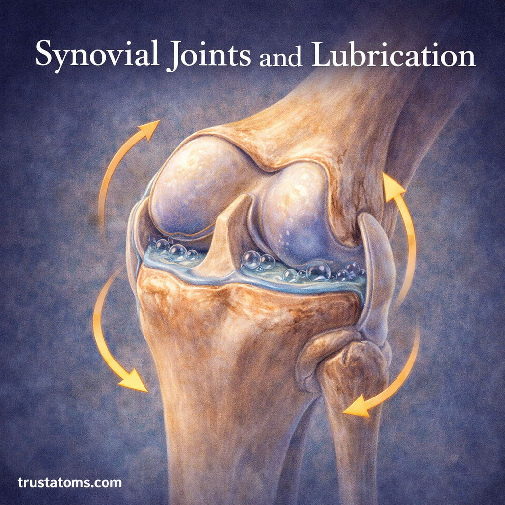
Synovial Joints and Lubrication

Illustration of a synovial knee joint showing cartilage surfaces and synovial fluid lubricating joint movement.
Diagram of a synovial joint demonstrating how fluid lubrication reduces friction and supports smooth movement. trustatoms.com.

Synovial joints are the most mobile and commonly used joints in the human body. From walking and running to lifting and rotating, these joints allow for smooth, controlled movement.

A key factor behind this efficiency is lubrication, which reduces friction and protects joint structures during motion. Understanding how synovial joints work—and how they stay lubricated—reveals why joint health is essential for everyday function.

Continue reading “Synovial Joints and Lubrication”

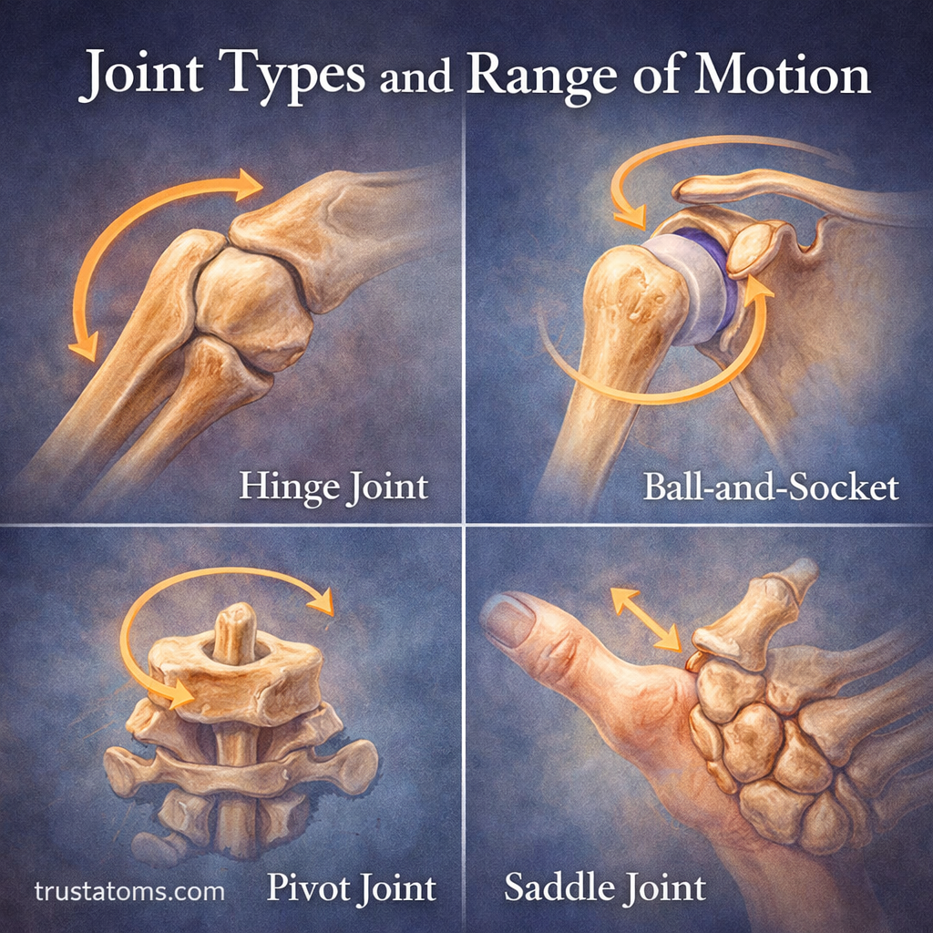
Joint Types and Range of Motion

Illustration showing different joint types including hinge, ball-and-socket, pivot, and saddle joints with their range of motion.
Different joint shapes permit specific movements, from bending at hinge joints to rotation and multidirectional movement at ball-and-socket joints.

Joints are connections between bones that provide stability, permit movement, or accomplish both functions. Their structure determines whether they remain nearly immovable, allow limited flexibility, or support a wide range of movements such as bending, rotating, and reaching.

Studying joints is a fundamental part of human anatomy because the skeleton must support body weight while allowing coordinated movement. Understanding joint types and their range of motion helps explain how the human body performs both simple and complex movements.

Continue reading “Joint Types and Range of Motion”

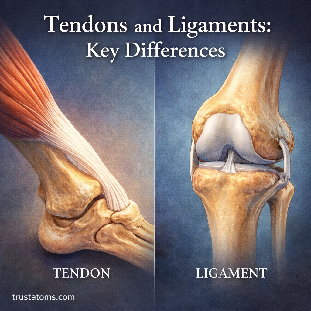
Tendons and Ligaments: Key Differences

Split illustration comparing a tendon connecting muscle to bone and a ligament connecting bone to bone at a joint.
Side-by-side comparison showing how tendons connect muscle to bone and ligaments connect bone to bone. trustatoms.com.

Tendons and ligaments are essential components of the musculoskeletal system, helping your body move, stabilize, and function efficiently. While they are often confused due to their similar structure and roles, they serve distinct purposes.

Understanding the difference between tendons and ligaments can help you better grasp how movement works—and why certain injuries occur.

Continue reading “Tendons and Ligaments: Key Differences”

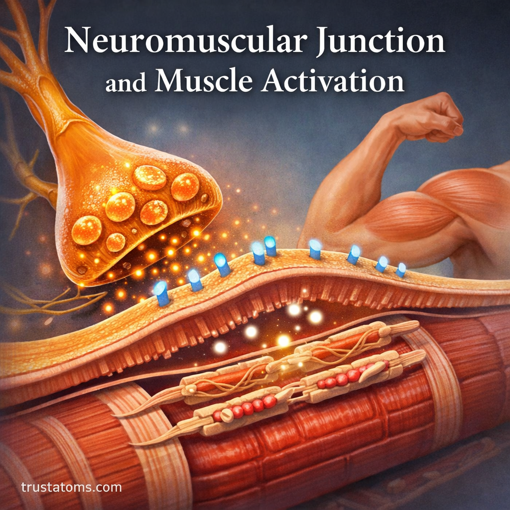
Neuromuscular Junction and Muscle Activation

Illustration of the neuromuscular junction showing a motor neuron releasing neurotransmitters to activate a muscle fiber.
Visual diagram of how nerve signals trigger muscle contraction at the neuromuscular junction. trustatoms.com.

The ability to move—whether lifting a weight, walking, or even blinking—relies on a precise connection between the nervous system and muscles. This connection occurs at a specialized site called the neuromuscular junction (NMJ).

Understanding how the neuromuscular junction works helps explain how muscles contract, how signals travel through the body, and what happens when this system is disrupted.

Continue reading “Neuromuscular Junction and Muscle Activation”

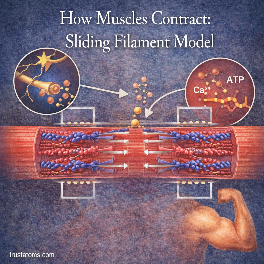
How Muscles Contract: Sliding Filament Model

Diagram illustrating the sliding filament model showing actin and myosin interaction during muscle contraction with ATP and calcium involvement.
Illustration of the sliding filament model explaining how muscles contract through actin and myosin interaction.

Muscles contract when myosin proteins repeatedly pull thin actin filaments toward the centers of microscopic units called sarcomeres, producing force through the sliding filament mechanism. The actin and myosin filaments do not shorten themselves; instead, their overlap changes as calcium regulates their interaction and ATP supplies energy for repeated movement.

This process allows skeletal muscles to generate the forces needed for walking, lifting, maintaining posture, and breathing. Understanding muscle contraction connects microscopic cell biology with human anatomy, showing how structures too small to see without a microscope produce visible movement.

The sliding filament model describes how individual muscle fibers generate force, but whole-muscle movement also depends on nerve signals, muscle fiber recruitment, and the mechanical load being moved. These factors determine whether a muscle shortens, stays the same length, or lengthens while producing tension.

Continue reading “How Muscles Contract: Sliding Filament Model”

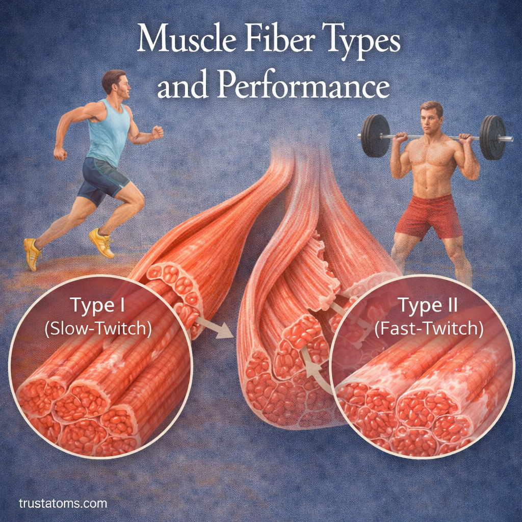
Muscle Fiber Types and Performance

Illustration comparing slow-twitch and fast-twitch muscle fibers with examples of endurance running and weightlifting.
Comparison of muscle fiber types showing how slow-twitch and fast-twitch fibers affect performance. trustatoms.com.

Not all muscles perform the same way. Some are built for endurance, while others are designed for speed and power. These differences come down to muscle fiber types.

Understanding muscle fiber types can help explain athletic performance, training adaptations, and why some people naturally excel at certain physical activities.

Continue reading “Muscle Fiber Types and Performance”

Skeletal Muscle Structure and Function

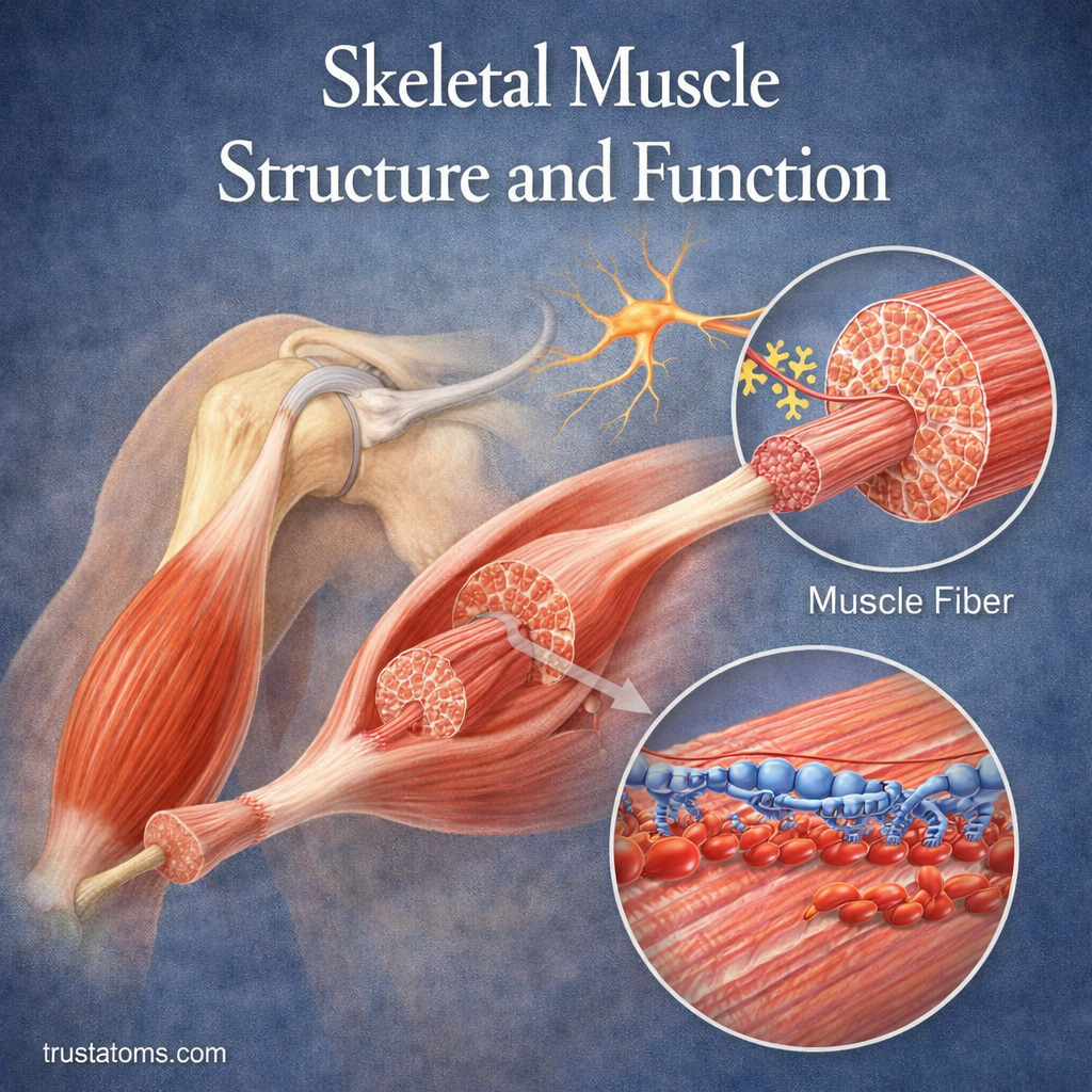
Illustration of skeletal muscle structure showing muscle fibers, fascicles, and actin-myosin interaction during contraction.
Diagram of skeletal muscle structure and function highlighting muscle fibers and contraction mechanism. trustatoms.com.

Skeletal muscles are the muscles attached to your bones that allow voluntary movement. Whether you’re walking, lifting, or even smiling, skeletal muscles are responsible for making those actions possible.

They are one of three muscle types in the body and play a key role in movement, posture, and overall physical strength.

Continue reading “Skeletal Muscle Structure and Function”

Bone Remodeling and Mineral Balance

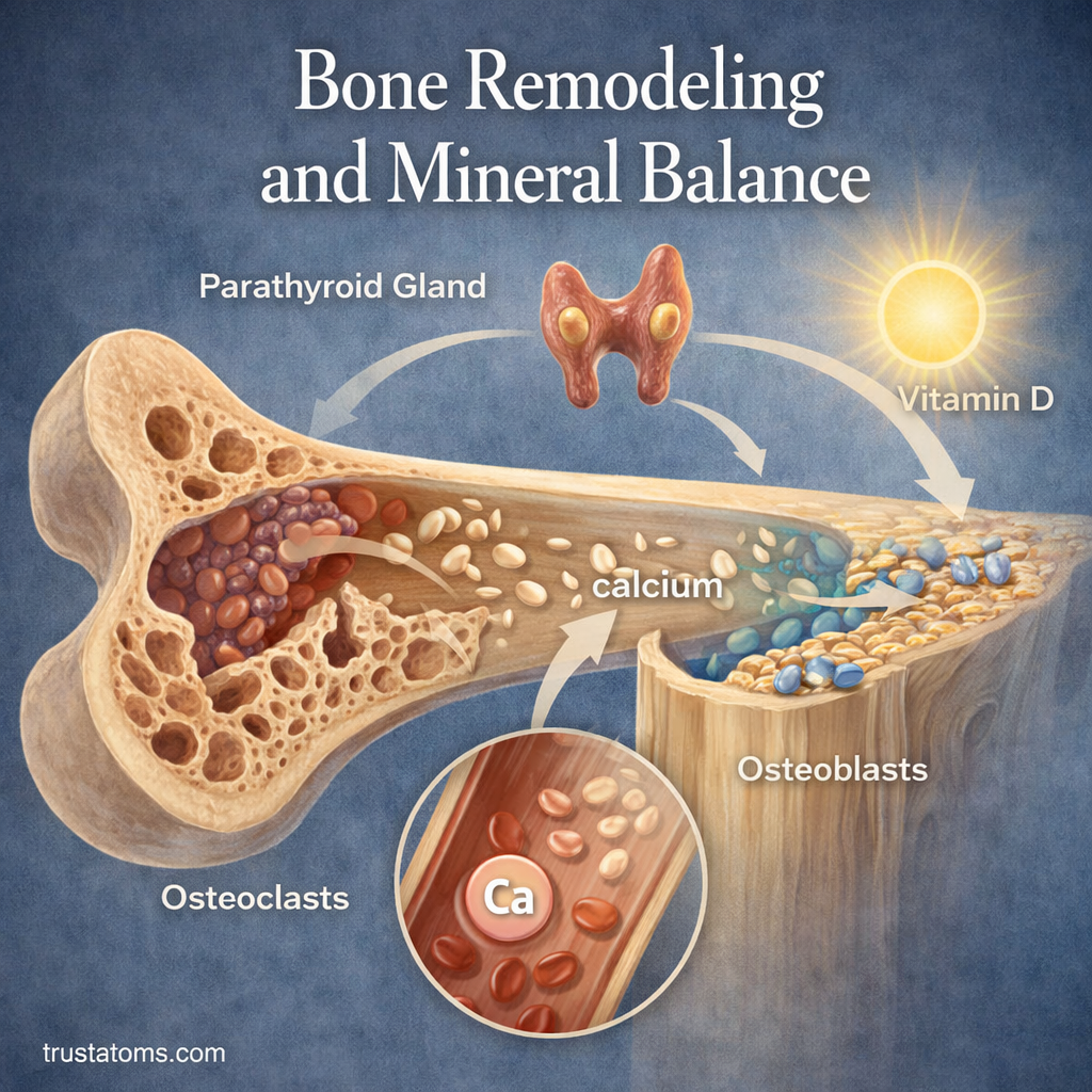
Illustration showing bone remodeling process with osteoclasts breaking down bone and osteoblasts rebuilding bone, highlighting calcium balance.
Osteoclasts remove existing bone while osteoblasts form new tissue, helping renew the skeleton and regulate stored minerals.

Bone remodeling is the continuous process of removing old bone tissue and replacing it with new bone, while mineral balance regulates how calcium and phosphorus are stored and used. Together, these processes help maintain skeletal strength, repair microscopic damage, and keep essential mineral concentrations within healthy ranges.

In human anatomy, bones are living organs rather than rigid structures that remain unchanged after growth. Their cells respond to mechanical forces, hormonal signals, and the body’s mineral requirements throughout life.

Continue reading “Bone Remodeling and Mineral Balance”

Bone Structure: Compact vs Spongy Bone

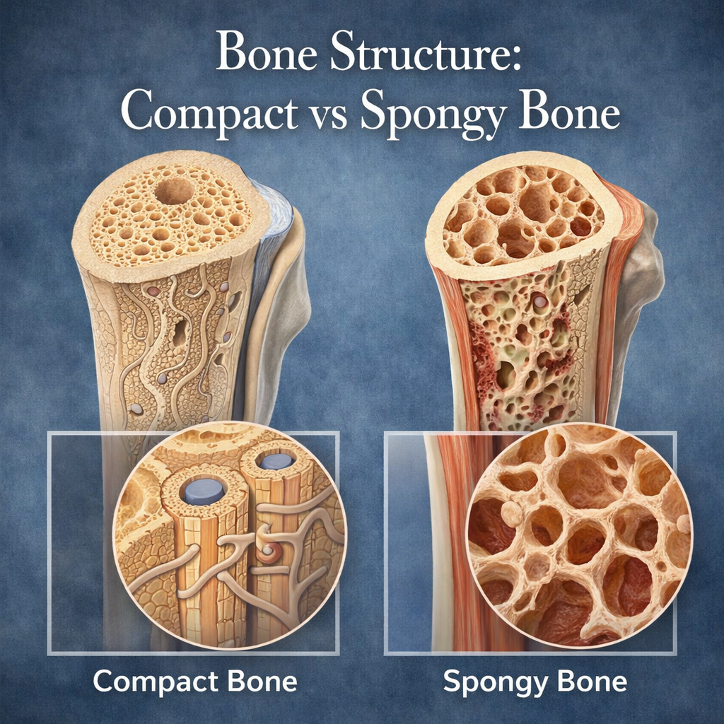
Diagram comparing compact bone and spongy bone showing dense outer bone and porous inner bone structure.
Comparison of compact and spongy bone structure highlighting density and internal composition.

Compact bone is the dense, strong tissue that forms the outer layer of bones, while spongy bone is a lighter, porous tissue found mainly inside them. Both are living bone tissues, but their different microscopic structures allow the skeleton to combine strength, efficient weight distribution, and space for bone marrow.

Although bones appear solid from the outside, their interiors contain organized networks of mineralized tissue, blood vessels, and living cells. Understanding these arrangements is an essential part of human anatomy because bone structure influences movement, organ protection, blood cell production, and the skeleton’s response to physical stress.

The main difference is architectural: compact bone has a tightly organized structure, while spongy bone consists of interconnected plates and struts called trabeculae. These tissues are not competing designs. They work together within individual bones to provide support without unnecessary weight.

Continue reading “Bone Structure: Compact vs Spongy Bone”