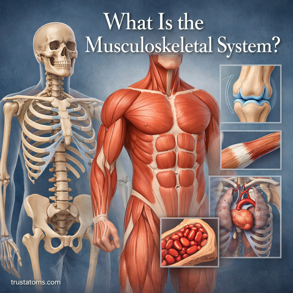
What Is the Musculoskeletal System?

Illustration showing the human musculoskeletal system with skeleton, muscles, joints, and connective tissues.
Illustration of the human musculoskeletal system highlighting bones, muscles, and joints.

The musculoskeletal system is the body’s structural and movement system. It includes bones, muscles, cartilage, tendons and ligaments, and joints working together to support your body, enable movement, and protect internal organs.

In human anatomy, the musculoskeletal system combines the skeleton’s rigid framework with muscles that generate force. Bones provide support and act as levers, while muscles pull on them through tendons to produce controlled movements.

Everyday activities such as walking, climbing stairs, lifting groceries, and maintaining posture depend on this coordination. The system also stores important minerals and contains bone marrow that produces blood cells.

Continue reading “What Is the Musculoskeletal System?”
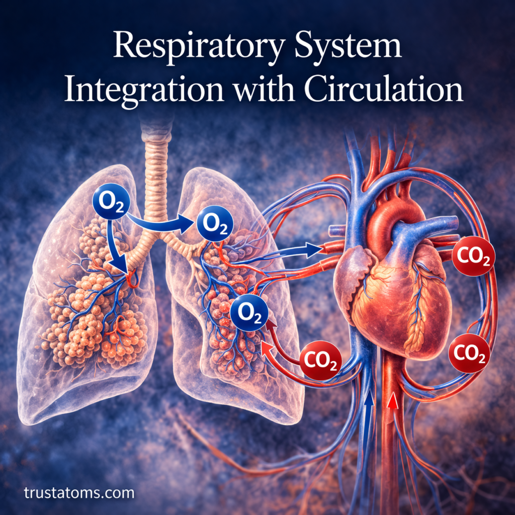
Respiratory System Integration with Circulation

Illustration showing how oxygen and carbon dioxide move between the lungs and heart in the respiratory and circulatory systems.
Illustration demonstrating the integration of the respiratory and circulatory systems, showing oxygen delivery and carbon dioxide removal. trustatoms.com

The human body relies on a tightly coordinated partnership between the respiratory and circulatory systems. While the lungs bring oxygen into the body, it’s the bloodstream that delivers that oxygen to every cell—and removes carbon dioxide in return.

This integration is essential for survival, energy production, and overall physiological balance. In this article, we’ll explore how these two systems work together to support life.

Continue reading “Respiratory System Integration with Circulation”

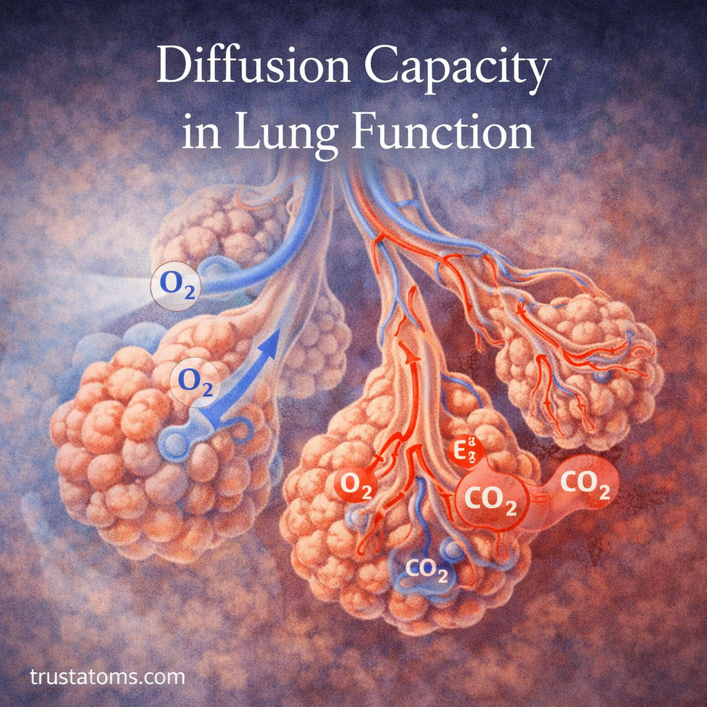
Diffusion Capacity in Lung Function

Illustration of gas exchange in the lungs, showing oxygen entering the blood and carbon dioxide leaving through the alveoli. trustatoms.com
Illustration of gas exchange in the lungs, showing oxygen entering the blood and carbon dioxide leaving through the alveoli.

Diffusion capacity describes the ability of the lungs to transfer gas from the alveoli into the bloodstream across the alveolar-capillary barrier. It depends on the available exchange surface, membrane properties, pulmonary capillary blood volume, and the blood’s ability to bind the transferred gas.

Breathing brings oxygen into the lungs, but oxygen must still cross a thin tissue barrier before entering the circulation. This connection between human anatomy and lung physiology helps explain why some respiratory conditions impair oxygen transfer even when air can enter and leave the lungs.

Continue reading “Diffusion Capacity in Lung Function”

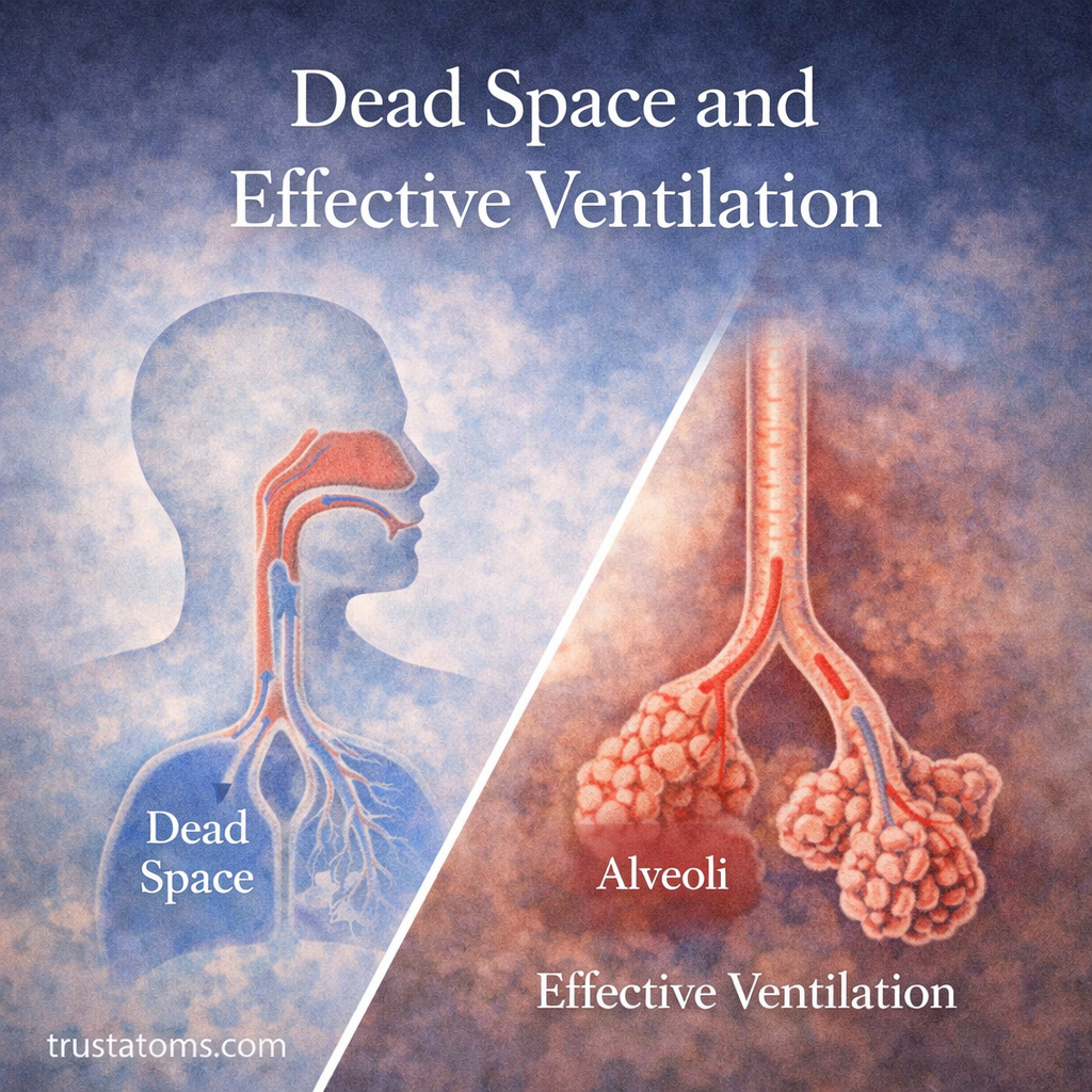
Dead Space and Effective Ventilation

Illustration showing dead space in the upper airways and effective ventilation in the alveoli of the lungs.
Illustration comparing dead space in the respiratory tract with effective ventilation occurring in the alveoli.

Respiratory dead space is the portion of inhaled air that does not participate in oxygen and carbon dioxide exchange. Effective ventilation, also called alveolar ventilation, describes how much fresh air reaches the gas-exchanging regions of the lungs each minute.

Not every milliliter of air entering the respiratory tract reaches functioning alveoli. Understanding this distinction connects human anatomy with respiratory physiology and explains why breathing depth, lung blood flow, and respiratory disease affect how efficiently the body exchanges gases.

Continue reading “Dead Space and Effective Ventilation”

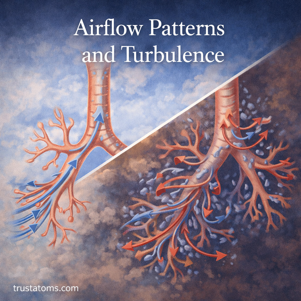
Airflow Patterns and Turbulence

Illustration comparing laminar and turbulent airflow patterns in the human respiratory system.
Illustration showing the difference between smooth laminar airflow and chaotic turbulent airflow in the respiratory system.

Airflow in the respiratory system follows two main patterns: laminar flow, in which air moves smoothly in organized layers, and turbulent flow, in which air moves irregularly with swirling currents. The pattern depends on airflow speed, airway dimensions, shape, and the physical properties of air, influencing how much pressure is needed to move air into and out of the lungs.

These patterns connect the physics of moving gases with human anatomy. Understanding them helps explain why breathing becomes more demanding during exercise, why narrowed airways restrict airflow, and how certain respiratory sounds develop.

Continue reading “Airflow Patterns and Turbulence”

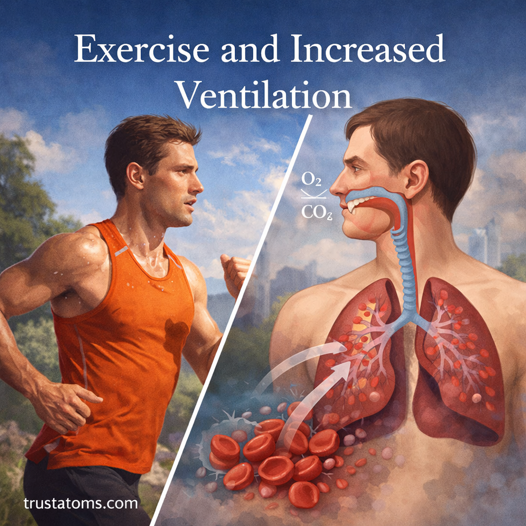
Exercise and Increased Ventilation

Illustration of a runner with a split view of the respiratory system showing oxygen intake and carbon dioxide release during exercise.
Illustration showing how exercise increases ventilation, with faster oxygen intake and carbon dioxide removal in the lungs.

Exercise increases ventilation because working muscles consume more oxygen and produce more carbon dioxide as their energy demands rise. The respiratory system responds by increasing breathing depth and frequency, allowing the lungs to exchange more gases while helping maintain normal blood chemistry.

Even a brisk walk can make breathing noticeably deeper, while running may require much faster breaths. Understanding this response connects human anatomy with the physiology of the lungs, respiratory muscles, circulation, and exercise performance.

Continue reading “Exercise and Increased Ventilation”

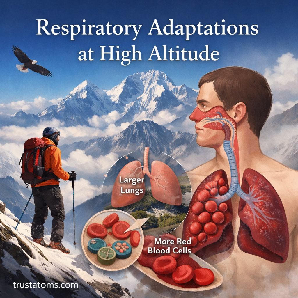
Respiratory Adaptations at High Altitude

Illustration of respiratory adaptations at high altitude showing a mountaineer, lungs, and increased red blood cells.
Illustration of how the human respiratory system adapts to high altitude with increased lung function and oxygen transport. trustatoms.com

High-altitude environments present a unique challenge to the human body. As elevation increases, atmospheric pressure drops, meaning less oxygen is available with each breath. Despite this, millions of people live, work, and even perform intense physical activity at high altitudes.

So how does the body adapt?

This article explores the fascinating respiratory changes that occur when humans are exposed to high-altitude conditions—whether for a short trip or long-term living.

Continue reading “Respiratory Adaptations at High Altitude”

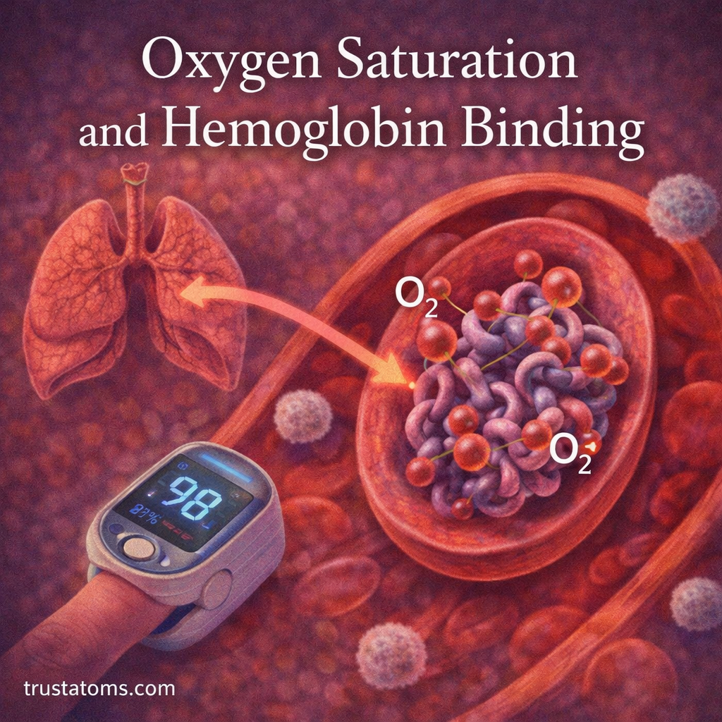
Oxygen Saturation and Hemoglobin Binding

Illustration showing oxygen binding to hemoglobin in red blood cells with lungs and a pulse oximeter indicating oxygen saturation.
Oxygen binds to hemoglobin in red blood cells, allowing efficient transport from the lungs to body tissues. trustatoms.com.

Oxygen is essential for life, powering the body’s cells and enabling energy production. But oxygen doesn’t travel freely in large amounts through the blood—it relies on a specialized protein called hemoglobin to be transported efficiently.

The relationship between oxygen and hemoglobin determines oxygen saturation, a key indicator of how well oxygen is being delivered throughout the body.

Continue reading “Oxygen Saturation and Hemoglobin Binding”

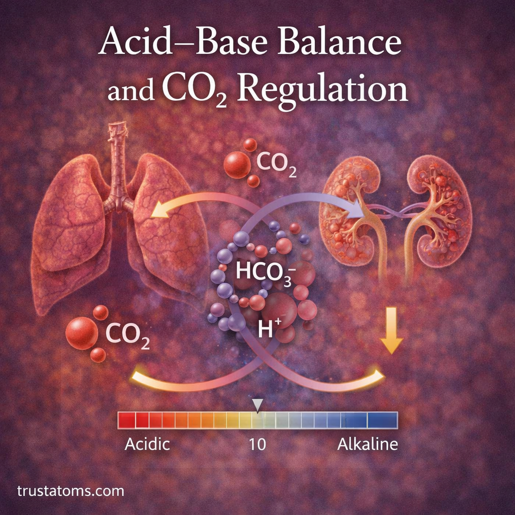
Acid–Base Balance and CO₂ Regulation

Illustration showing lungs and kidneys regulating carbon dioxide and pH balance through bicarbonate and hydrogen ion exchange.
The lungs and kidneys work together to regulate CO2 levels and maintain acid–base balance in the body.

Acid–base balance is the process of maintaining the body’s hydrogen ion concentration within a narrow range so cells and organs can function normally. Carbon dioxide (CO₂) helps regulate this balance because changes in its concentration affect blood acidity, while the lungs and kidneys work together to stabilize pH.

In human anatomy, acid–base regulation connects the respiratory, circulatory, and urinary systems. The lungs adjust how much CO₂ leaves the body, while the kidneys regulate bicarbonate and hydrogen ions to provide longer-term control.

These adjustments occur continuously, including during exercise, sleep, and changes in breathing. They help keep arterial blood pH near its normal range of 7.35–7.45.

Continue reading “Acid–Base Balance and CO₂ Regulation”

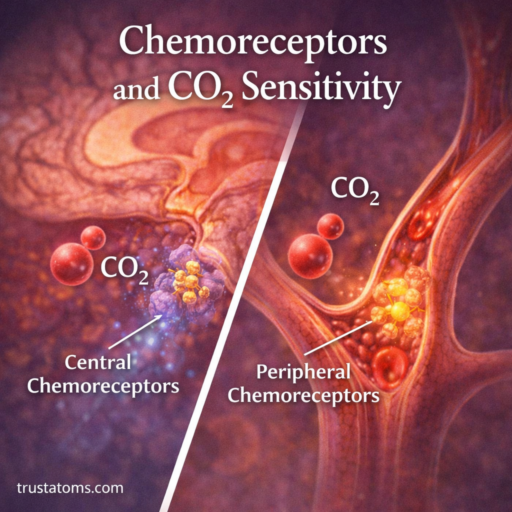
Chemoreceptors and CO₂ Sensitivity

Illustration showing central chemoreceptors in the brain and peripheral chemoreceptors in arteries detecting carbon dioxide levels.
Chemoreceptors detect changes in carbon dioxide levels and signal the brain to adjust breathing rate and depth.

Chemoreceptors are specialized sensory cells and neural structures that detect chemical changes related to carbon dioxide (CO₂), oxygen, and acidity, helping the body regulate breathing automatically. CO₂ sensitivity describes how strongly the respiratory control system responds to changes in carbon dioxide, usually by adjusting the depth and frequency of breaths.

This feedback system connects the lungs, blood vessels, and brainstem, making it an important example of how structures studied in human anatomy work together. It helps keep blood gases and acid-base conditions within ranges compatible with normal cellular function, even as activity levels change.

Continue reading “Chemoreceptors and CO₂ Sensitivity”