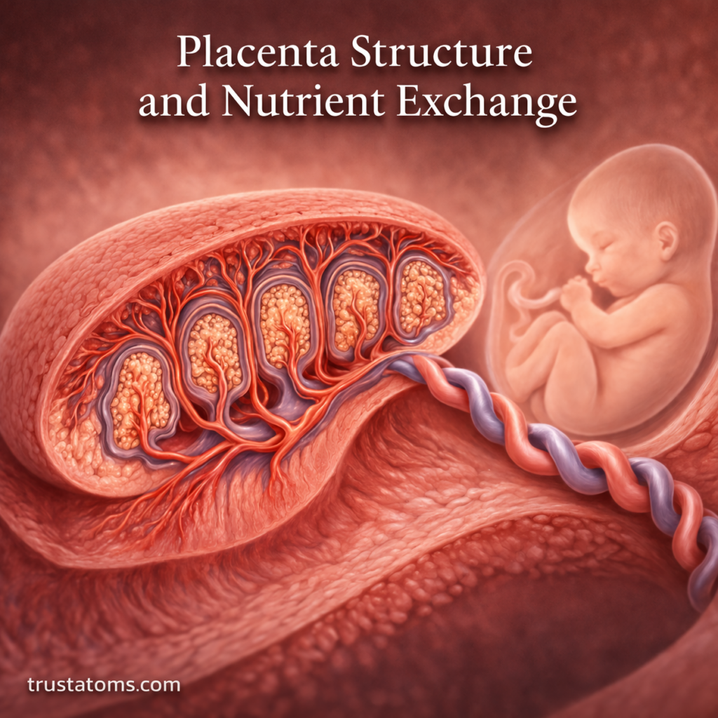
Placenta Structure and Nutrient Exchange

Simple illustration of placenta structure showing umbilical cord connection and nutrient exchange between mother and fetus.
Clean diagram of placenta structure and nutrient exchange during pregnancy. trustatoms.com

The placenta is a remarkable organ that forms during pregnancy to support the developing embryo and fetus. It acts as a lifeline between the mother and the baby, enabling the exchange of nutrients, oxygen, and waste products. Understanding the structure of the placenta and how nutrient exchange occurs provides key insights into human development and maternal health.

Continue reading “Placenta Structure and Nutrient Exchange”
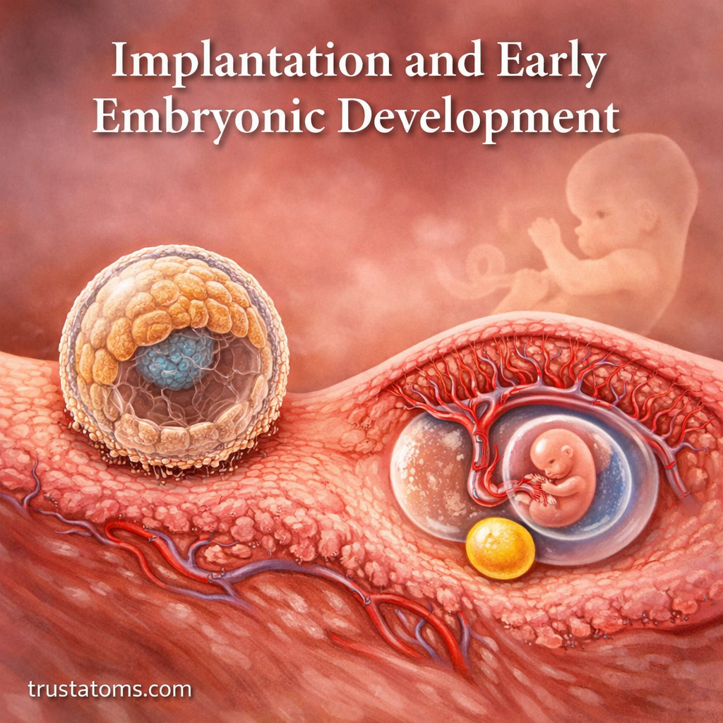
Implantation and Early Embryonic Development

Illustration showing implantation of a blastocyst into the uterine lining and early embryonic development stages.
A blastocyst attaches to the uterine lining and begins developing the structures needed to support early pregnancy.

Implantation occurs when a blastocyst, an early stage of the developing embryo, attaches to and embeds within the uterine lining. Early embryonic development then establishes the cell layers and supporting structures that eventually form the body’s organs and tissues.

These events begin within days of fertilization and involve coordinated changes in the embryo and uterus. Understanding their timing connects reproductive biology with human anatomy, showing how a single fertilized cell develops into increasingly specialized structures.

Continue reading “Implantation and Early Embryonic Development”

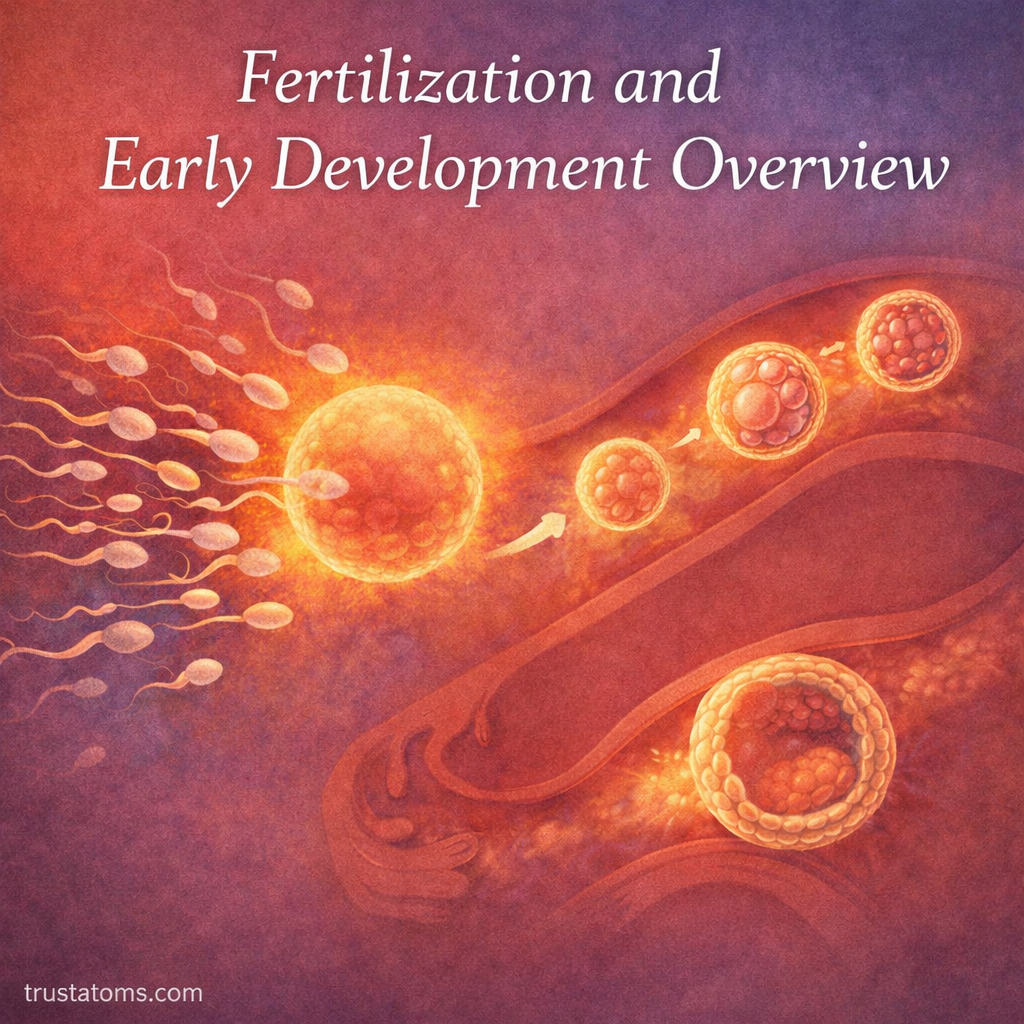
Fertilization and Early Development Overview

Illustration showing fertilization and early development stages from sperm meeting egg to blastocyst formation.
Diagram illustrating fertilization and early embryonic development from zygote to blastocyst.

Fertilization occurs when a sperm cell fuses with an egg, initiating the formation of a zygote that begins dividing into increasingly specialized cells. During the following days, the developing embryo travels toward the uterus, forms a blastocyst, and undergoes a series of rapid changes as the newly formed organism prepares for implantation in the uterus.

These early events connect reproductive physiology, genetics, and human anatomy. Understanding their sequence helps explain how pregnancy begins, why timing matters, and how the reproductive tract supports development before an embryo becomes established in the uterine lining.

Continue reading “Fertilization and Early Development Overview”

Ovulation and Fertilization Processes

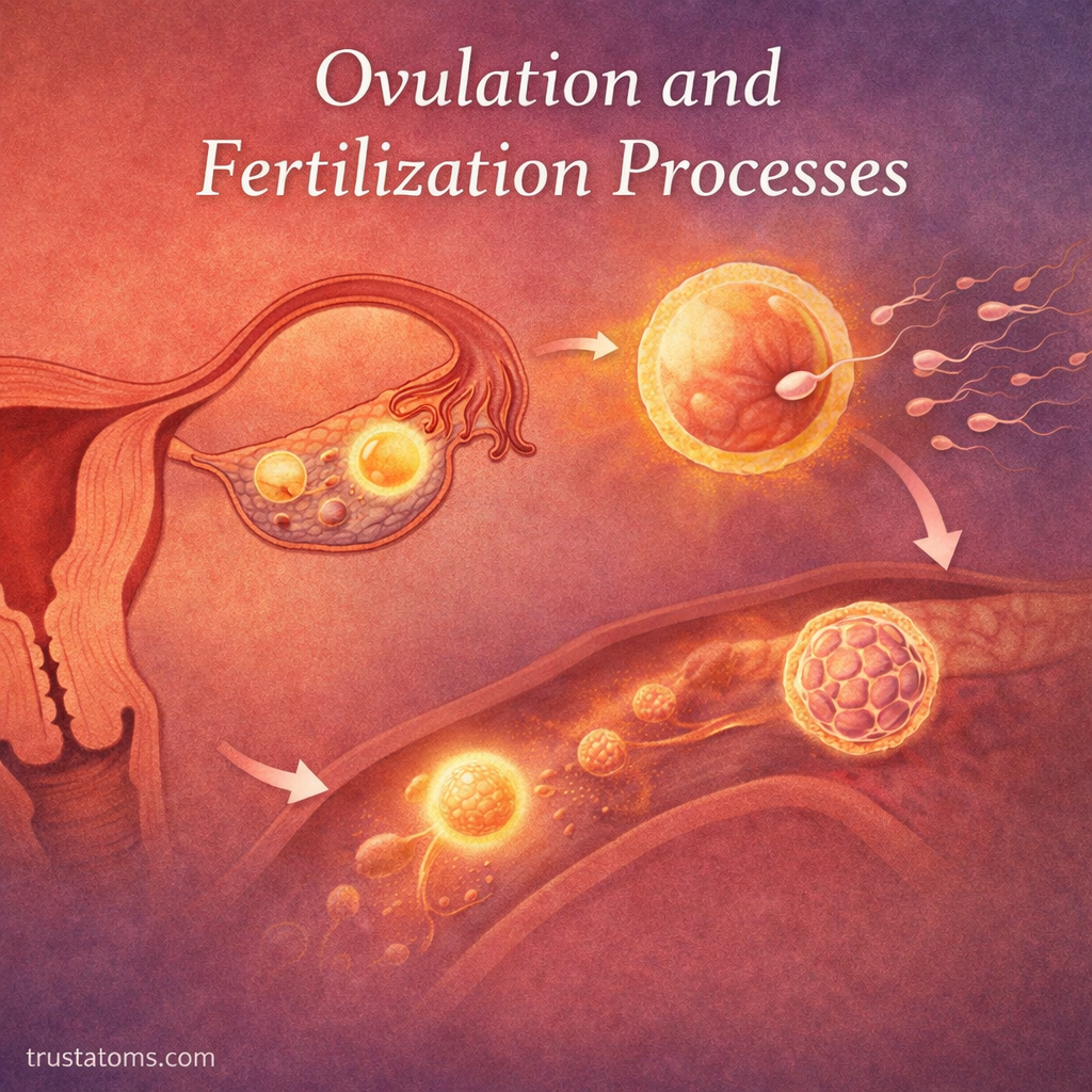
Illustration showing ovulation and fertilization processes including egg release, sperm movement, and early embryo development.
Diagram illustrating the journey from ovulation to fertilization and early embryo development. trustatoms.com

Ovulation and fertilization are central events in human reproduction. These processes involve the release of an egg from the ovary and its potential union with sperm, leading to the formation of a new life.

Understanding how ovulation and fertilization work provides insight into fertility, reproductive health, and the biological timing required for conception.

Continue reading “Ovulation and Fertilization Processes”

Menstrual Cycle Phases and Hormonal Control

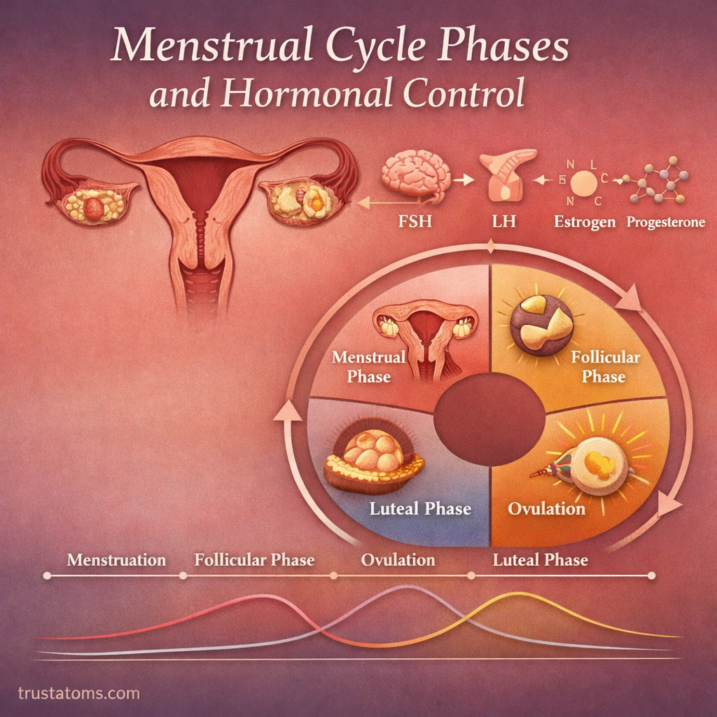
Illustration showing menstrual cycle phases and hormonal control including menstrual, follicular, ovulation, and luteal phases.
Simple diagram of the menstrual cycle phases and how hormones regulate each stage. trustatoms.com

The menstrual cycle is a coordinated series of physiological changes in the female body that prepares it for potential pregnancy. Controlled by a complex interaction of hormones, the cycle typically lasts about 28 days (though 21–35 days is normal for adults).

Understanding the phases of the menstrual cycle and the hormones that regulate them is essential for learning about reproductive health, fertility, and overall hormonal balance.

Continue reading “Menstrual Cycle Phases and Hormonal Control”

Oogenesis in Female Reproduction

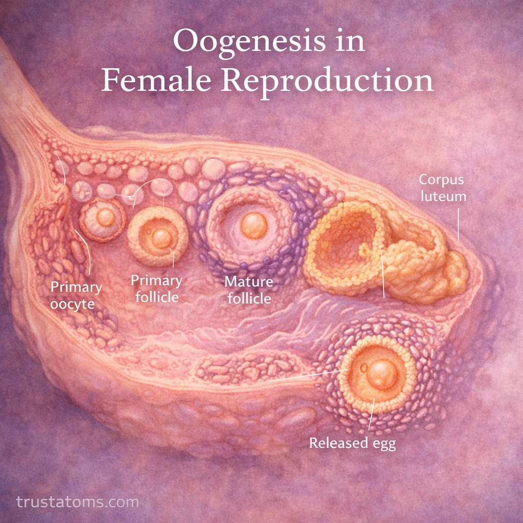
Diagram of oogenesis showing egg development stages in the ovary from primary oocyte to released egg.
Illustration showing oogenesis and the development of egg cells in the ovary. trustatoms.com

Oogenesis is the biological process through which female reproductive cells, known as eggs (ova), are produced. This process occurs in the ovaries and is essential for human reproduction, as it generates the cells required for fertilization.

Unlike sperm production, oogenesis is a cyclical and finite process that begins before birth and continues until menopause.

Continue reading “Oogenesis in Female Reproduction”

Spermatogenesis in Male Reproduction

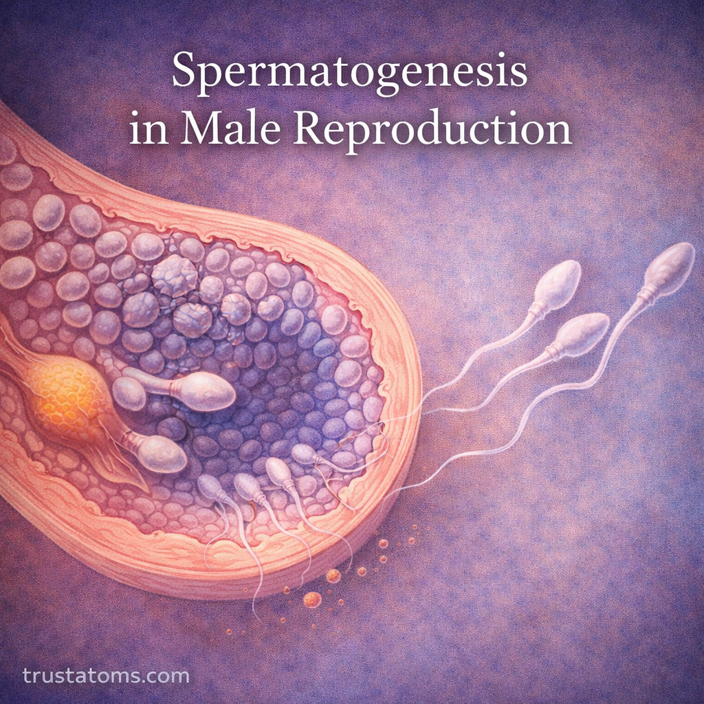
Diagram of spermatogenesis showing sperm development inside seminiferous tubules in the testes.
Illustration showing spermatogenesis and the development of sperm cells in the testes. trustatoms.com

Spermatogenesis is the biological process by which male reproductive cells—sperm—are produced. This process occurs in the testes and is essential for human reproduction, as it generates the cells needed to fertilize an egg.

Spermatogenesis is continuous, highly regulated, and begins at puberty, continuing throughout a male’s life.

Continue reading “Spermatogenesis in Male Reproduction”

Gametes and Reproductive Cells

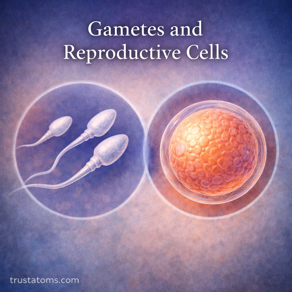
Illustration of gametes showing sperm cells and an egg cell representing male and female reproductive cells.
Illustration showing sperm and egg cells as the primary human gametes involved in reproduction.

Gametes are specialized reproductive cells that carry half the usual number of chromosomes and combine during fertilization to form a new organism. In humans, sperm and egg cells each contribute 23 chromosomes, allowing the resulting zygote to normally contain 46 chromosomes.

Although sperm and eggs differ greatly in size, structure, and development, both are essential to sexual reproduction. Studying these cells connects human anatomy with genetics, cell division, and the earliest stages of human development.

Continue reading “Gametes and Reproductive Cells”

Female Reproductive Anatomy Overview

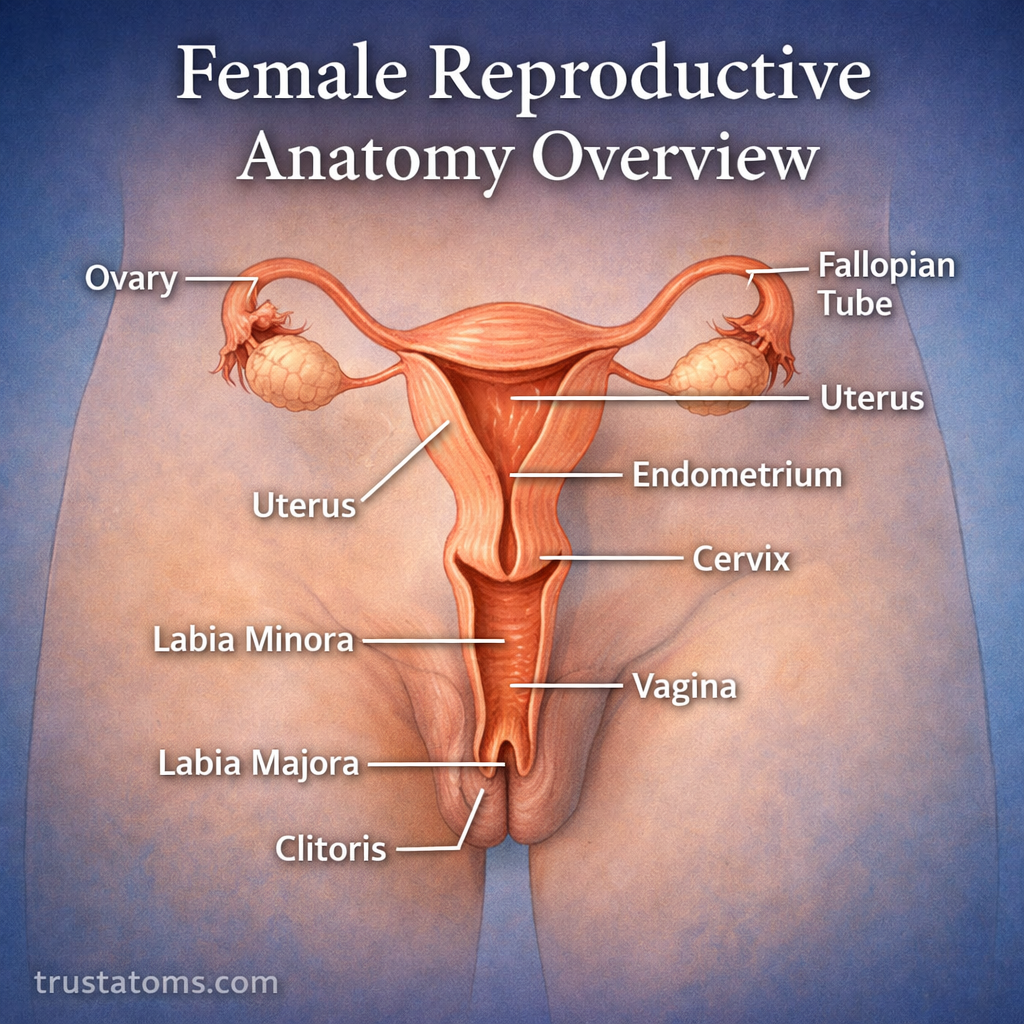
Female reproductive anatomy overview diagram showing labeled organs including ovaries, fallopian tubes, uterus, cervix, vagina, and vulva structures.
Illustrated overview of female reproductive anatomy highlighting key internal and external organs.

The female reproductive system is a complex and highly coordinated group of organs responsible for producing eggs, supporting fertilization, and nurturing the development of a fetus. Its main structures include the ovaries, fallopian tubes, uterus, cervix, vagina, and vulva, which work together through hormonal signals and specialized anatomical functions.

Understanding this reproductive system connects the study of human anatomy with menstruation, fertility, pregnancy, and reproductive health. Each organ has a distinct structure, but reproductive processes depend on their coordinated activity.

Continue reading “Female Reproductive Anatomy Overview”

Male Reproductive Anatomy Overview

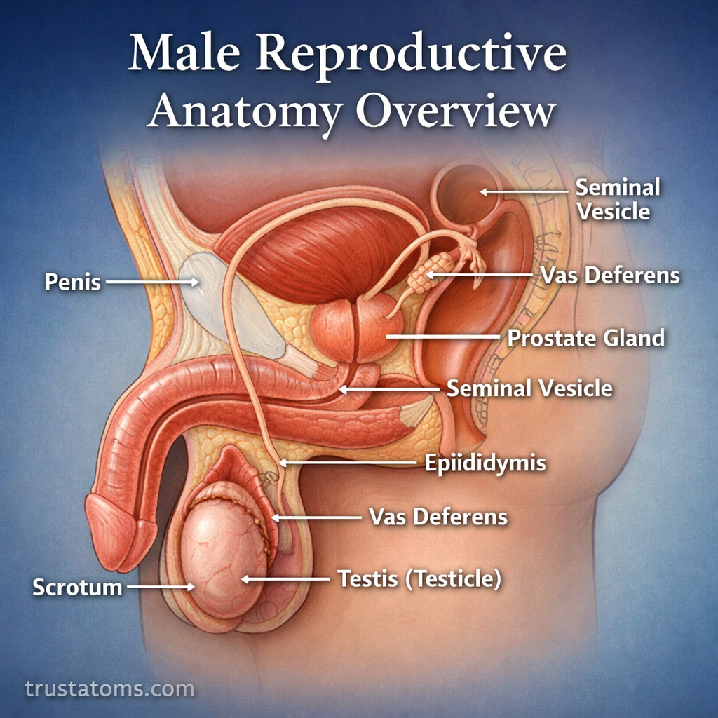
Male reproductive anatomy overview diagram showing labeled internal and external organs including penis, testes, epididymis, vas deferens, prostate, and seminal vesicles.
Illustrated overview of male reproductive anatomy highlighting key organs and their structure. trustatoms.com

The male reproductive system is a specialized group of organs responsible for producing, storing, and delivering sperm, as well as generating male sex hormones like testosterone. These structures work together to support reproduction, sexual function, and overall hormonal balance.

Understanding male reproductive anatomy helps explain how fertility works, how hormones regulate the body, and how different organs contribute to the reproductive process.

Continue reading “Male Reproductive Anatomy Overview”