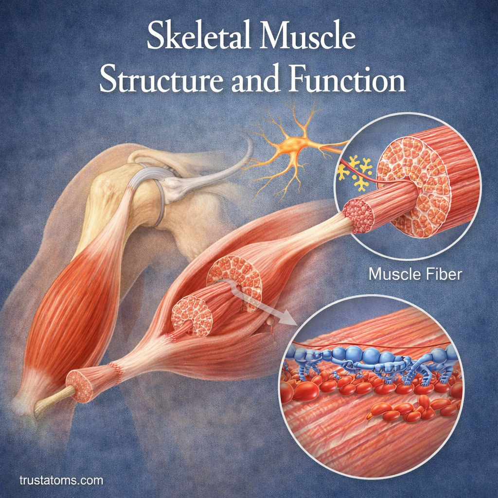
Skeletal muscle is a type of striated muscle tissue that produces body movement by generating force through the interaction of actin and myosin proteins. Its organized structure, from whole muscles to microscopic sarcomeres, allows the body to walk, lift objects, maintain posture, and stabilize joints.
Most skeletal muscles attach to bones through tendons and receive signals from motor neurons. Understanding their structure and function connects muscle physiology with human anatomy and explains how the nervous and skeletal systems cooperate during movement.
What Is Skeletal Muscle?
Skeletal muscle is one of three major muscle tissue types, alongside cardiac and smooth muscle. Its cells have a striped appearance under a microscope because their contractile proteins are arranged in repeating patterns.
Unlike cardiac and most smooth muscle, skeletal muscle is generally under voluntary control. It works with bones and joints as part of the Musculoskeletal System.
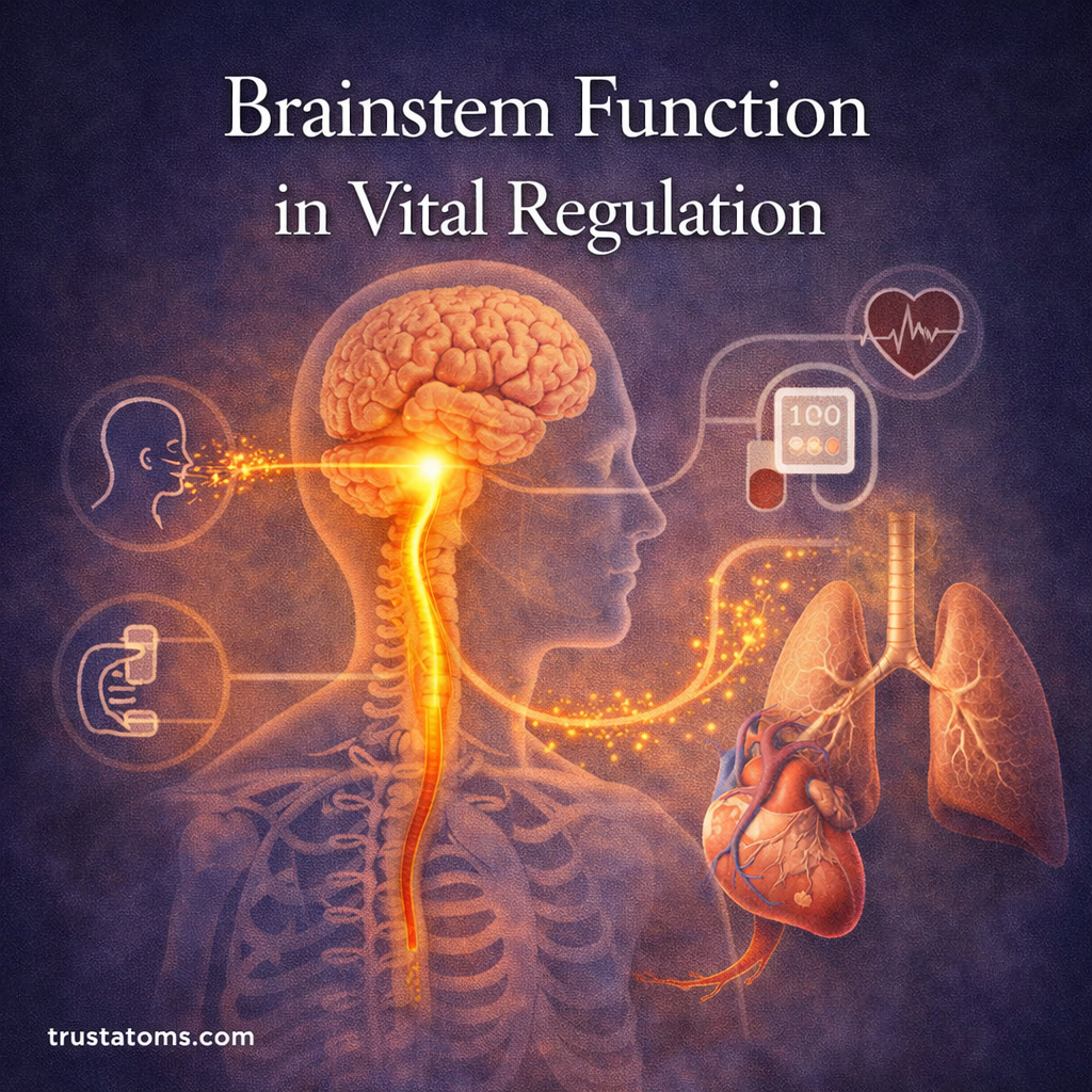
Motor neurons activate skeletal muscle fibers, allowing the nervous system to control contraction strength and timing. Somatic Nervous System in Voluntary Control explains how these signals connect the brain, spinal cord, and muscles.
How Skeletal Muscle Is Organized
Skeletal muscles have a layered organization that allows thousands of microscopic contractile structures to work together.
- Whole muscle: The complete organ, such as the biceps brachii, surrounded by connective tissue called epimysium.
- Fascicles: Bundles of muscle fibers enclosed by perimysium.
- Muscle fibers: Long, multinucleated muscle cells individually surrounded by endomysium.
- Myofibrils: Contractile structures extending through muscle fibers.
- Sarcomeres: Repeating functional units containing organized actin and myosin filaments.
The connective tissue layers help transmit muscular force toward tendons and bones. Blood vessels supply oxygen and nutrients, while nerves deliver signals that activate contraction.
Muscle Fibers, Myofibrils, and Sarcomeres
A skeletal muscle fiber is a specialized cell containing many nuclei, mitochondria, and myofibrils. Its cell membrane is called the sarcolemma, and its internal fluid is the sarcoplasm.
Each myofibril contains sarcomeres arranged end to end. A sarcomere extends between two Z discs and contains thin filaments made primarily of actin and thick filaments made primarily of myosin.
During contraction, myosin pulls actin filaments toward the sarcomere’s center. The filaments themselves do not shorten; instead, their overlap increases, shortening the sarcomere.
When many sarcomeres generate force simultaneously, their combined action produces tension throughout the muscle. Depending on the external load, the whole muscle may shorten, lengthen under tension, or remain approximately the same length.
How Skeletal Muscles Work
Muscle contraction begins when a motor neuron sends an electrical signal to a muscle fiber at a specialized connection called the neuromuscular junction.
- Nerve activation: The motor neuron releases acetylcholine, triggering an electrical impulse in the muscle fiber.
- Signal transmission: The impulse spreads along the sarcolemma and into transverse tubules.
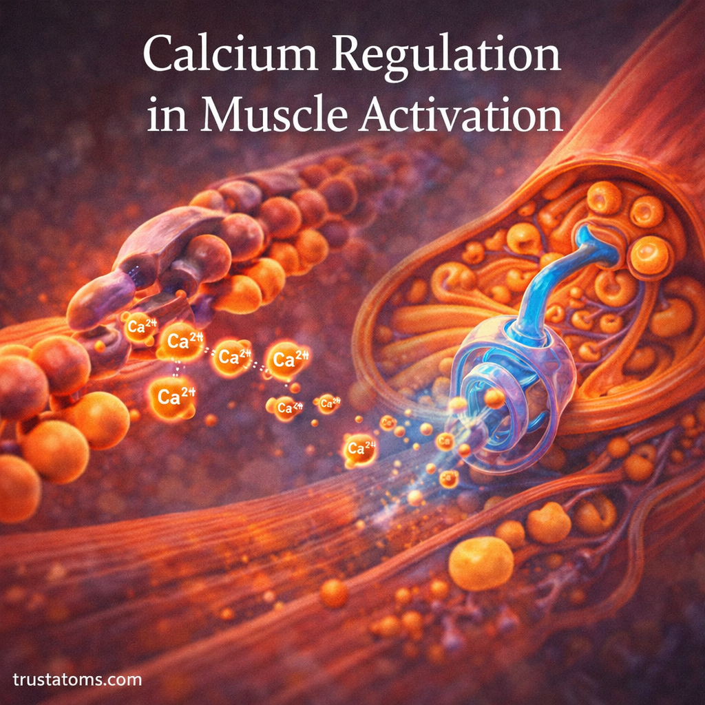
- Calcium release: The sarcoplasmic reticulum releases calcium ions into the muscle cell.
- Cross-bridge cycling: Calcium allows myosin heads to bind actin and generate force using ATP.
- Relaxation: Calcium is pumped back into the sarcoplasmic reticulum, reducing actin-myosin interactions.
This process is called excitation-contraction coupling. The molecular details of filament movement are explained in How Muscles Contract.
Slow-Twitch and Fast-Twitch Muscle Fibers
Skeletal muscle fibers differ in contraction speed, force production, and resistance to fatigue. Most muscles contain a mixture of fiber types.
| Fiber Type | Main Characteristics | Typical Activities |
|---|---|---|
| Type I | Slow contraction, high fatigue resistance, strong aerobic capacity | Posture and sustained endurance activity |
| Type IIa | Faster contraction, relatively high force, moderate fatigue resistance | Repeated powerful movements |
| Type IIx | Very fast contraction, high power potential, lower fatigue resistance | Brief explosive efforts |
Fiber characteristics depend partly on genetics, training, and the muscle’s function. Endurance and resistance training can change muscle performance and some fiber properties, although training does not freely convert every fiber into any other type.
How Muscles Control Movement in Pairs
Skeletal muscles pull on bones rather than push them. Because a muscle generates tension in one direction, coordinated movement often requires muscles with opposing actions.
- Agonist: The muscle primarily responsible for producing a particular movement.
- Antagonist: The muscle that opposes the movement.
- Synergist: A muscle that assists the movement or helps control unwanted motion.
For example, when lifting a cup toward the mouth, the biceps brachii contributes to elbow flexion while the triceps brachii can regulate opposing tension. When straightening the elbow, their primary movement roles reverse.
Antagonists do not always become completely inactive. Controlled co-contraction can help stabilize joints, particularly during precise or demanding movements.
Energy Use in Muscle Contraction
Muscle fibers require adenosine triphosphate (ATP) to generate force, release myosin from actin, and pump calcium during relaxation.
Because stored ATP is limited, muscles continuously regenerate it through several overlapping energy systems.
- Phosphocreatine system: Rapidly regenerates ATP during short, intense efforts.
- Glycolysis: Breaks down glucose or glycogen to produce ATP, including when oxygen availability is limited.
- Oxidative metabolism: Uses oxygen to generate ATP from carbohydrates, fats, and other fuels during sustained activity.
These pathways operate together, but their relative contributions change with exercise intensity and duration. ATP Use in Muscle Contraction explains the energy requirements of the cross-bridge cycle.
Functions Beyond Voluntary Movement
Skeletal muscles support several physiological functions in addition to moving the limbs.
- Posture: Continuous low-level muscle activity helps maintain body position.
- Joint stability: Muscles generate controlled forces that support joint alignment.
- Heat production: Muscle metabolism produces heat, including during shivering.
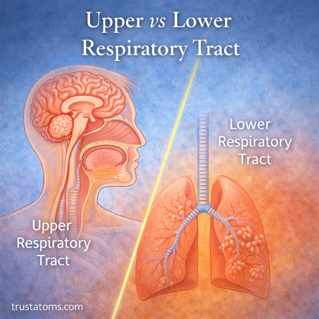
- Breathing: The diaphragm and intercostal muscles produce movements necessary for ventilation.
- Metabolic regulation: Skeletal muscle stores glycogen and contributes substantially to glucose uptake.
Common Skeletal Muscle Injuries and Conditions
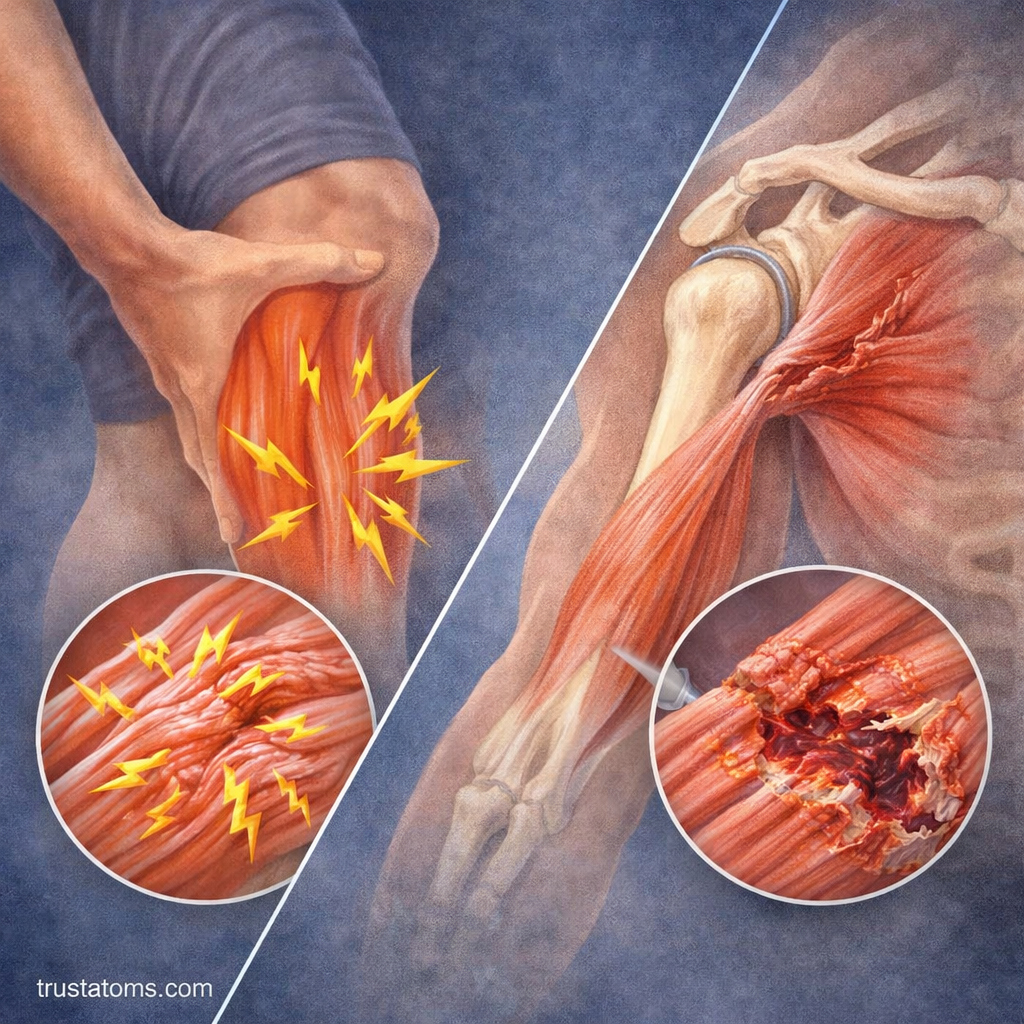
Muscles can be affected by temporary functional disturbances, physical injuries, or diseases involving muscle tissue and its nerve supply.
- Muscle strains: Overstretching or excessive loading can damage muscle fibers or nearby connective tissue.
- Muscle cramps: Sudden, involuntary contractions that may be painful and can have several contributing causes.
- Muscular dystrophies: Inherited disorders that cause progressive muscle weakness and degeneration.
- Muscle atrophy: Reduction in muscle size associated with inactivity, aging, illness, or impaired nerve supply.
Tendinopathy can also affect the tendons that transmit muscular force. Persistent weakness, significant pain, or unexplained muscle loss should be medically evaluated.
Maintaining Skeletal Muscle Health
Regular physical activity helps preserve muscle strength, endurance, and functional capacity. Resistance exercise encourages muscle adaptation, while aerobic activity supports cardiovascular fitness and muscle metabolism.
Adequate dietary protein supplies amino acids needed for tissue maintenance and repair. Sufficient energy intake, hydration, sleep, and recovery also support normal muscular function.
Training loads should increase gradually according to individual ability. Recovery requirements vary with exercise intensity, health, and previous training experience.
Frequently Asked Questions
What is the basic functional unit of skeletal muscle?
The sarcomere is the basic contractile unit of skeletal muscle. It contains organized actin and myosin filaments that interact to generate force.
What makes skeletal muscle different from smooth muscle?
Skeletal muscle has a striated appearance, long multinucleated fibers, and generally operates under voluntary control. Smooth muscle lacks visible striations and typically controls involuntary movements in organs and blood vessels.
Do skeletal muscles always shorten when they contract?
No. Muscles can shorten during concentric contractions, lengthen while generating tension during eccentric contractions, or maintain nearly constant length during isometric contractions.
Skeletal muscle converts electrical signals and chemical energy into mechanical force through highly organized contractile structures. From individual sarcomeres to coordinated muscle groups, this system supports movement, posture, breathing, and joint stability. Its performance depends on functioning nerves, adequate energy supplies, and the ability of muscle tissue to adapt to physical demands.
This article is for general education and is not medical advice. Talk to a qualified health professional about any medical concern.
Sources: Guyton and Hall Textbook of Medical Physiology; Principles of Anatomy and Physiology by Tortora and Derrickson; OpenStax Anatomy and Physiology 2e.