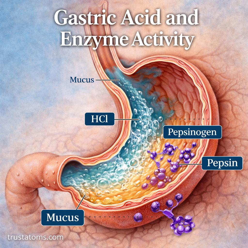
Gastric acid and digestive enzymes work together in the stomach to unfold food proteins and break them into smaller peptides. Hydrochloric acid creates the acidic environment needed to activate pepsin, while muscular contractions mix food with gastric juices and prepare it for further digestion in the small intestine.
This coordinated process illustrates how human anatomy supports chemical digestion through specialized cells, protective tissues, and carefully regulated secretions. Although most nutrient absorption occurs in the small intestine, the stomach prepares food for the digestive processes that follow.
What Is Gastric Acid?
Gastric acid is a component of gastric juice secreted by specialized cells in the stomach lining. Its main acidic substance is hydrochloric acid (HCl), which produces the low pH needed for several digestive functions.
Gastric juice in an empty stomach commonly has a pH of approximately 1–3, although acidity varies with meals, secretion rates, and other conditions. Because the pH scale is logarithmic, a decrease of one pH unit represents a tenfold increase in hydrogen ion concentration.
The stomach is not only a muscular organ for mixing food—it is also a chemical environment where protein digestion begins. The distinction between these processes is explored in Mechanical vs Chemical Digestion.
After food leaves the stomach, pancreatic bicarbonate helps neutralize the acidic material entering the duodenum. Pancreas Function in Digestion explains how this neutralization supports enzyme activity in the small intestine.
Which Stomach Cells Produce Acid and Enzymes?
The stomach lining contains gastric glands with specialized cells that produce different components of gastric juice.
| Cell Type | Main Secretion | Function |
|---|---|---|
| Parietal cells | Hydrochloric acid and intrinsic factor | Create acidity and support vitamin B12 absorption |
| Chief cells | Pepsinogen and gastric lipase | Supply digestive enzyme precursors and enzymes |
| Surface mucous cells | Mucus and bicarbonate | Protect the stomach lining |
| G cells | Gastrin | Stimulate gastric acid secretion |
| Enterochromaffin-like cells | Histamine | Stimulate parietal cells |
These secretions work together, allowing the stomach to digest food while protecting its own tissues from acid and enzymes.
How Gastric Acid Helps Digest Food
Hydrochloric acid contributes to digestion in several ways. Its effects extend beyond simply making stomach contents acidic.
- Protein denaturation: Acid disrupts interactions that maintain protein structure, causing proteins to unfold and become more accessible to enzymes.
- Enzyme activation: Acid promotes the conversion of inactive pepsinogen into active pepsin.
- Microbial defense: Strong acidity inactivates many microorganisms entering with food, although some pathogens can survive.
- Nutrient preparation: Acidity helps release certain nutrients from food and supports the solubility of some minerals, including non-heme iron.
Stomach acid also helps release vitamin B12 from food proteins. However, absorption of vitamin B12 occurs later in the ileum and requires intrinsic factor produced by parietal cells.
Pepsin and Gastric Lipase: The Main Stomach Enzymes
Pepsin Breaks Proteins Into Smaller Peptides
Pepsin is the primary enzyme responsible for protein digestion in the stomach. Chief cells release it as an inactive precursor called pepsinogen, helping prevent enzyme activity inside the cells that produce it.
In the acidic stomach environment, pepsinogen is converted into pepsin. Active pepsin then helps activate additional pepsinogen molecules and breaks peptide bonds within proteins.
Pepsin produces smaller peptides rather than completing protein digestion into individual amino acids. Most subsequent protein breakdown occurs in the small intestine through pancreatic and intestinal enzymes.
Gastric Lipase Begins Fat Digestion
Gastric lipase is another enzyme released by chief cells. It breaks some triglycerides into smaller lipid components and contributes to the early stages of fat digestion.
Its contribution is particularly relevant in infants, although it also functions in adults. Most dietary fat digestion occurs later in the small intestine with pancreatic lipase and bile salts.
Different digestive enzymes operate under different chemical conditions, which explains why stomach and intestinal digestion require distinct environments.
How Gastric Acid Activates Pepsin During Digestion
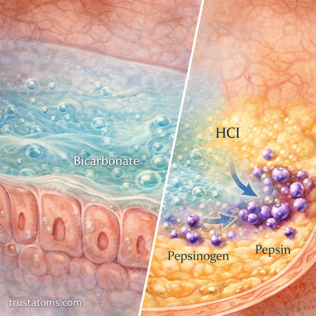
Acid secretion, enzyme activation, and muscular mixing occur together after food enters the stomach.
- Food enters the stomach and stimulates mechanical and chemical signals.
- Parietal cells secrete hydrochloric acid into the stomach lumen.
- Chief cells release pepsinogen into gastric juice.
- The acidic environment activates pepsinogen, producing pepsin.
- Pepsin breaks protein molecules into smaller peptides while stomach contractions mix the contents.
- Partially digested food, called chyme, gradually passes through the pylorus into the duodenum.
A Practical Example: Digesting a Protein-Rich Meal
Imagine eating a meal containing chicken, rice, and vegetables. Chewing breaks the food into smaller pieces, and swallowing moves it into the stomach.
Once the chicken reaches the stomach, hydrochloric acid helps unfold its proteins. Pepsin then cuts some of those proteins into shorter peptide chains while stomach contractions mix the meal with gastric juice.
As chyme enters the small intestine, pancreatic enzymes continue breaking the peptides into smaller fragments and amino acids that can be absorbed. This example shows why stomach digestion is an early stage of protein processing rather than the final step.
How the Body Regulates Gastric Acid Secretion
Acid secretion is regulated through neural, hormonal, and local chemical signals. Physiologists commonly describe three overlapping phases.
- Cephalic phase: Seeing, smelling, tasting, or anticipating food activates vagal signals that prepare the stomach for digestion.
- Gastric phase: Stomach stretching and the presence of peptides stimulate acid secretion through neural signals and gastrin release.
- Intestinal phase: Signals from the small intestine generally reduce gastric acid secretion and slow gastric emptying when needed.
Gastrin, histamine, and acetylcholine stimulate parietal cells. When stomach acidity becomes sufficiently high, somatostatin helps suppress gastrin release, limiting further acid production.
How the Stomach Protects Itself From Acid
The stomach lining must tolerate conditions that would damage many other tissues. Several defenses help prevent acid and pepsin from injuring its surface.
- Mucus: Forms a protective layer over the epithelial surface.
- Bicarbonate: Helps maintain a less acidic environment immediately next to surface cells.
- Tight junctions: Limit the movement of acid between neighboring epithelial cells.
- Blood flow and tissue repair: Support nutrient delivery, removal of damaging substances, and replacement of injured cells.
When these defenses are disrupted, the stomach becomes more vulnerable to inflammation and ulcer formation.
How pH Changes Gastric Enzyme Activity
Pepsin works most effectively in strongly acidic conditions, generally around pH 1.5–2. As pH rises, its activity decreases substantially, and it becomes inactive in near-neutral conditions.
After a meal, food temporarily buffers stomach acid and raises gastric pH. Continued acid secretion gradually restores a more acidic environment as digestion proceeds.
When chyme enters the duodenum, bicarbonate neutralizes much of its acidity. This protects intestinal tissues and creates conditions suitable for pancreatic enzymes.
Common Problems Involving Gastric Acid
Several digestive disorders involve gastric acid, impaired protective mechanisms, or abnormal exposure of other tissues to stomach contents.
- Gastroesophageal reflux disease (GERD): Stomach contents repeatedly flow into the esophagus, causing symptoms or tissue injury. Reflux does not necessarily mean excessive acid production.
- Peptic ulcers: Sores in the stomach or duodenal lining, commonly associated with Helicobacter pylori infection or nonsteroidal anti-inflammatory drugs.
- Hypochlorhydria: Reduced stomach acid production that can interfere with certain digestive and nutrient-related processes.
- Gastritis: Inflammation of the stomach lining, which may result from infection, autoimmune disease, or other causes.
Persistent abdominal pain, repeated vomiting, difficulty swallowing, or unexplained weight loss warrants medical evaluation. Vomiting blood or passing black, tarry stools requires urgent assessment.
Frequently Asked Questions
Does stomach acid directly break down proteins?
Stomach acid mainly unfolds proteins and creates conditions that activate pepsin. Pepsin performs much of the enzymatic breakdown of proteins into smaller peptides within the stomach.
Why does the stomach not digest itself?
The stomach protects its lining with mucus, bicarbonate, tightly connected epithelial cells, and rapid tissue repair. These defenses limit direct exposure of vulnerable cells to acid and digestive enzymes.
What happens to pepsin when food enters the small intestine?
As acidic chyme is neutralized in the duodenum, the rising pH causes pepsin to lose activity. Pancreatic proteases then take over much of the remaining protein digestion.
Gastric acid creates the chemical conditions that allow pepsin to begin protein digestion, while stomach contractions expose food to these secretions. Hormonal feedback and protective mucus help balance digestive activity with tissue safety. Once chyme enters the small intestine, a less acidic environment allows pancreatic enzymes to continue nutrient breakdown.
This article is for general education and is not medical advice. Talk to a qualified health professional about any medical concern.
Sources: Guyton and Hall Textbook of Medical Physiology; Boron and Boulpaep Medical Physiology; National Institute of Diabetes and Digestive and Kidney Diseases (NIDDK).