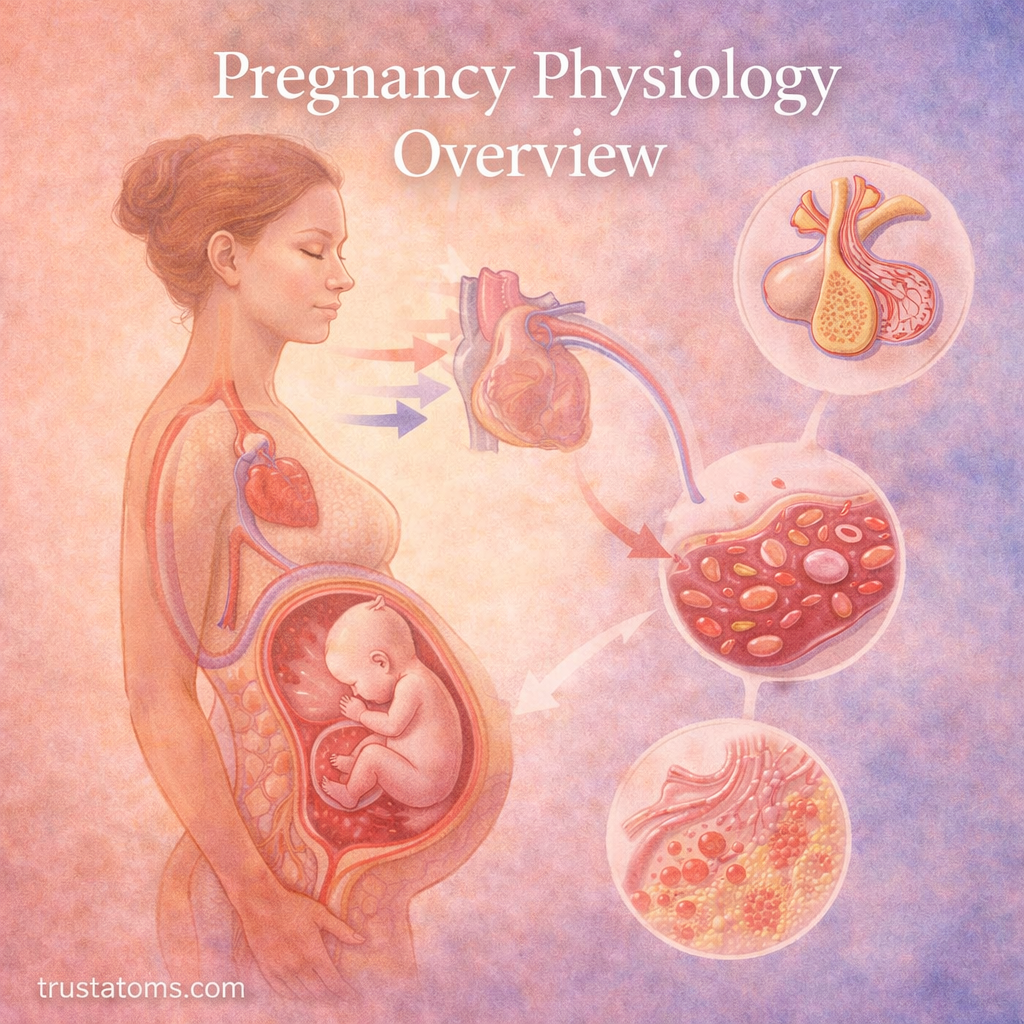
Pregnancy physiology describes the hormonal, anatomical, and functional changes that occur in the body during pregnancy. These adaptations support fetal growth, increase oxygen and nutrient delivery, regulate maternal metabolism, and prepare the uterus and other organs for childbirth.
Pregnancy affects far more than the reproductive system. Understanding these changes through human anatomy and physiology reveals how the cardiovascular, respiratory, digestive, renal, and endocrine systems adjust together throughout gestation.
What Is Pregnancy Physiology?
Pregnancy physiology examines how the maternal body maintains pregnancy while meeting the changing demands of a developing embryo and fetus. These adaptations begin early, sometimes before pregnancy is recognized, and continue through delivery.
The uterus, ovaries, cervix, and surrounding structures provide the anatomical foundation for pregnancy. The female reproductive anatomy overview explains how these structures support fertilization, implantation, and gestation.
Hormonal communication coordinates many of these changes. Understanding reproductive hormones and regulation helps explain how the menstrual cycle transitions into the hormonal environment required to maintain pregnancy.
Hormonal Changes During Pregnancy
Pregnancy depends on hormones produced by the ovaries, placenta, and maternal endocrine organs. Their concentrations and effects change as gestation progresses.
| Hormone | Main Function During Pregnancy |
|---|---|
| Human chorionic gonadotropin (hCG) | Maintains the corpus luteum early in pregnancy, supporting progesterone production. |
| Progesterone | Supports the uterine lining and helps maintain uterine relaxation. |
| Estrogens | Support uterine growth, blood flow, and changes in maternal tissues. |
| Human placental lactogen (hPL) | Helps adjust maternal metabolism and nutrient availability. |
| Relaxin | Contributes to changes in reproductive tissues and connective tissue properties. |
| Prolactin | Supports mammary gland development and preparation for milk production. |
During early pregnancy, hCG helps preserve the corpus luteum. As the placenta develops, it increasingly produces progesterone and estrogens needed to maintain pregnancy.
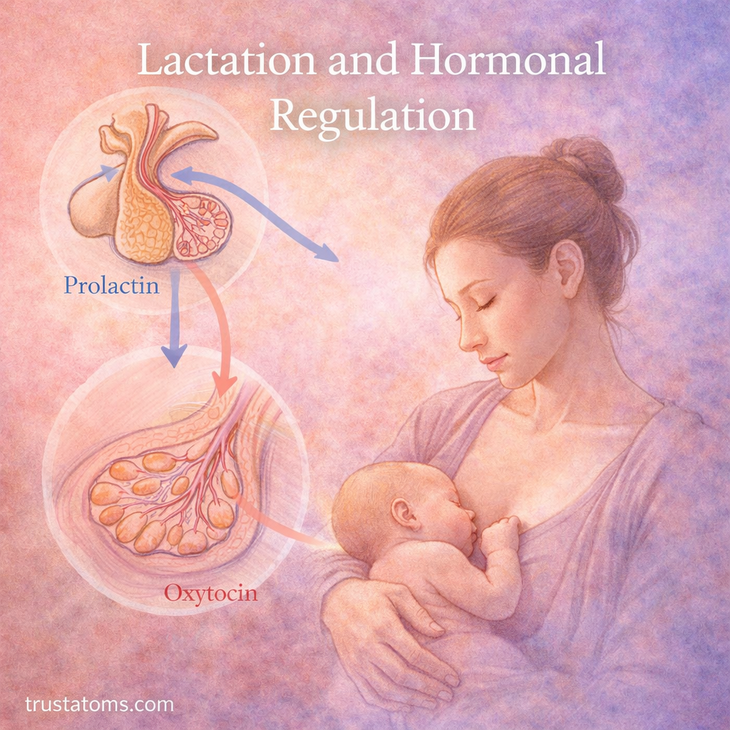
Breast tissue also changes in preparation for feeding after delivery. The relationship between prolactin, oxytocin, and milk production is explored in lactation and hormonal regulation.
Cardiovascular Changes: More Blood and Higher Cardiac Output
Pregnancy increases the demands placed on the heart and blood vessels. Maternal blood volume typically rises by approximately 30–50%, while heart rate and cardiac output also increase.
Cardiac output is the amount of blood the heart pumps each minute. It rises because the heart generally beats faster and pumps more blood per beat. Meanwhile, systemic vascular resistance decreases, partly because blood vessels relax and placental circulation develops.
Blood pressure may decrease during the middle of pregnancy before moving closer to prepregnancy levels later. Plasma volume also increases proportionally more than red blood cell mass, producing the normal dilutional effect sometimes called physiological anemia of pregnancy.
A Concrete Example of Blood Volume Expansion
Consider a person with approximately 5 liters of circulating blood before pregnancy. A 40% increase would bring the total to about 7 liters, an additional 2 liters. This is an illustrative calculation rather than a prediction for an individual pregnancy, since blood volume changes vary with body size and other factors.
The expanded circulation helps supply the uterus and placenta, supports maternal tissues, and provides a physiological reserve for blood loss during delivery.
Respiratory Changes: Breathing More Air
Pregnancy increases oxygen consumption and changes breathing patterns. Progesterone stimulates the respiratory system, increasing ventilation primarily by raising tidal volume, the amount of air moved with each breath.
Breathing rate usually changes little. Instead, deeper breaths increase the amount of air entering and leaving the lungs each minute. This lowers maternal carbon dioxide levels and helps support carbon dioxide transfer from fetal to maternal circulation.
As the uterus enlarges, the diaphragm moves upward and functional residual capacity decreases. Some pregnant people experience a greater awareness of breathing even when oxygen levels remain normal.
Digestive and Kidney Adaptations
Digestive System Changes
Hormonal changes influence gastrointestinal function throughout pregnancy. Nausea and vomiting are especially common early in gestation, while relaxation of smooth muscle and changes in digestive motility can contribute to constipation and acid reflux.
As the uterus grows, pressure on abdominal organs may increase reflux symptoms. Digestive experiences vary considerably, and not every pregnant person develops the same symptoms.
Kidney and Urinary Changes
Renal blood flow and glomerular filtration rate increase during pregnancy, allowing the kidneys to process a greater volume of blood. The kidneys also adjust sodium and water handling to support expanded maternal fluid volume.
Urinary frequency may increase because of hormonal influences and pressure from the enlarging uterus. Increased filtration does not necessarily mean urine production rises continuously, since the kidneys also retain additional fluid.
Uterine Growth and Placental Function
The uterus expands dramatically during pregnancy as its muscle cells enlarge and its tissues stretch. Uterine blood flow increases to support the developing placenta and fetus.
The placenta is a vital organ that connects mother and fetus. It forms an exchange interface between maternal and fetal circulations and also produces hormones that help maintain pregnancy.
- Gas exchange: Oxygen moves toward fetal blood while carbon dioxide moves toward maternal blood.
- Nutrient transfer: Glucose, amino acids, fatty acids, vitamins, and minerals cross through different transport mechanisms.
- Waste removal: Fetal metabolic waste products enter maternal circulation for elimination.
- Hormone production: Placental hormones influence maternal metabolism, uterine function, and breast development.
Maternal and fetal blood normally remain in separate circulatory compartments, although small amounts of cellular material may cross. The placenta is a selective exchange barrier, not an absolute shield against every substance or infection.
Fetal Development and Nutrient Exchange
The fetus receives oxygen and nutrients through placental exchange and the umbilical cord. The umbilical vein carries oxygenated, nutrient-rich blood toward the fetus, while two umbilical arteries return blood toward the placenta.
Fetal blood circulation differs from circulation after birth because the lungs are not yet responsible for oxygen exchange. The fetal circulation overview explains how specialized pathways direct blood through the developing body before the transition to breathing air.
Placental exchange depends on adequate maternal circulation, functioning placental tissue, and fetal blood flow. These interacting factors influence the delivery of nutrients needed for growth and organ development.
Immune and Musculoskeletal Changes
The maternal immune system undergoes carefully regulated changes during pregnancy. Rather than becoming uniformly suppressed, immune activity shifts in ways that support fetal tolerance while preserving defenses against many infections. These changes vary across tissues and stages of pregnancy.
Musculoskeletal adaptations occur as body mass and weight distribution change. The growing abdomen shifts the center of mass, which may alter standing posture, walking patterns, and balance. Changes in connective tissues and joint loading can also contribute to pelvic or lower back discomfort.
The Three Trimesters of Pregnancy
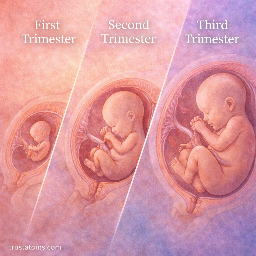
Pregnancy is commonly divided into three trimesters, with gestational age counted from the first day of the last menstrual period rather than the date of fertilization.
| Trimester | Major Developments |
|---|---|
| First (weeks 1–13) | Implantation, placental development, early organ formation, and substantial hormonal changes. |
| Second (weeks 14–27) | Continued fetal growth, movement becoming noticeable, and further organ development. |
| Third (week 28 to birth) | Rapid weight gain, continued brain and lung maturation, and preparation for delivery. |
Trimester boundaries can vary slightly between references. Development also occurs continuously, so these divisions are convenient descriptions rather than abrupt biological transitions.
Frequently Asked Questions
Why does blood volume increase during pregnancy?
Blood volume expands to support uterine and placental circulation, meet maternal metabolic demands, and provide additional circulatory reserve during childbirth.
Does the placenta mix maternal and fetal blood?
Normally, maternal and fetal blood circulate in separate compartments. Oxygen, nutrients, and waste products cross the placental exchange barrier without requiring the two blood supplies to mix directly.
When do physiological changes begin during pregnancy?
Changes begin very early, with hormonal signaling associated with implantation and maintenance of the corpus luteum. Cardiovascular, renal, respiratory, and metabolic adaptations then develop progressively as pregnancy continues.
Pregnancy physiology involves coordinated adjustments across nearly every major maternal body system. Hormonal regulation, expanded circulation, increased ventilation, and placental exchange work together to support fetal development while maintaining maternal function. These changes progress throughout gestation and gradually transition toward the physiological demands of childbirth and recovery.
This article is for general education and is not medical advice. Talk to a qualified health professional about any medical concern.
Sources: Williams Obstetrics; Guyton and Hall Textbook of Medical Physiology; Merck Manual Professional Edition.