Sexual differentiation is the developmental process through which an embryo forms reproductive structures and other physical characteristics associated with biological sex. It begins before birth and depends on coordinated genetic signals, gonadal development, hormone production, and the responsiveness of developing tissues.
Early embryos share several reproductive structures that can develop along different pathways. Understanding this process connects embryology with human anatomy and helps explain how reproductive organs form and why anatomical variations sometimes occur.
What Is Sexual Differentiation?
Sexual differentiation describes the formation of reproductive anatomy from initially similar embryonic tissues. It includes the development of gonads, internal reproductive ducts, and external genital structures.
These processes occur in stages rather than through a single event. Chromosomes influence gene activity, genes direct gonadal development, and hormones guide the growth or regression of particular structures.
Understanding sexual differentiation provides insight into how the human reproductive system forms and why variations can occur.
How Sex Chromosomes Influence Development
Chromosomal sex is usually established at fertilization. Most people have either an XX or XY sex chromosome pattern, although other chromosome combinations and variations also occur.
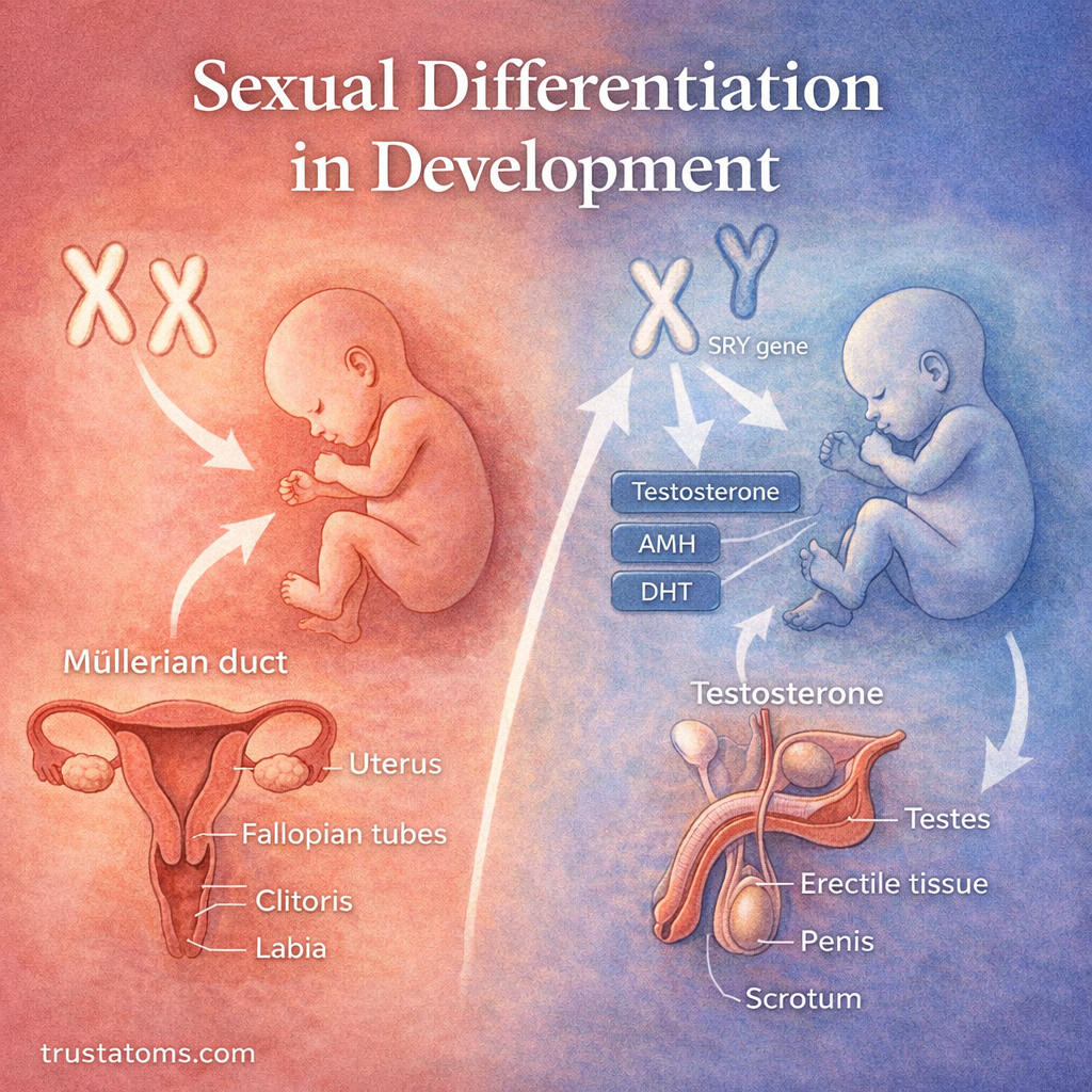
In typical XY development, the Y chromosome carries the SRY gene, which helps initiate testis formation. SRY activates a network of developmental genes, including SOX9, that supports differentiation of the initially undifferentiated gonad.
In typical XX development, ovarian formation depends on other gene networks, including pathways involving WNT4, RSPO1, and FOXL2. Ovarian development is therefore an actively regulated process, not simply the absence of testis development.
Chromosomes alone do not determine every anatomical outcome. Gene function, hormone production, and tissue responses all influence subsequent development.
The Early Embryo Has Shared Reproductive Structures
During early embryonic development, the gonads are initially bipotential, meaning they can develop into testes or ovaries depending on molecular signals.
The embryo also develops two paired reproductive duct systems:
- Mesonephric ducts: Also called Wolffian ducts, these can develop into several male internal reproductive structures.
- Paramesonephric ducts: Also called Müllerian ducts, these can form the uterine tubes, uterus, cervix, and upper portion of the vagina.
External genital tissues also begin with a similar arrangement, including the genital tubercle, urogenital folds, and labioscrotal swellings.
As development continues, genetic and hormonal signals direct these shared structures toward different anatomical outcomes.
Hormones That Guide Sexual Differentiation
Once the gonads begin differentiating, hormones influence the development of internal ducts and external genitalia. Three hormones are particularly important in typical male fetal development.
Testosterone
Fetal Leydig cells in the testes produce testosterone. This hormone supports the maintenance and differentiation of Wolffian ducts into structures such as the epididymis, vas deferens, and seminal vesicles.
Anti-Müllerian Hormone
Sertoli cells in developing testes produce anti-Müllerian hormone (AMH). This hormone causes the Müllerian ducts to regress during typical male development.
Dihydrotestosterone
Dihydrotestosterone (DHT) forms when the enzyme 5-alpha-reductase converts testosterone into a more potent androgen. DHT supports typical masculinization of the external genitalia and prostate development.
In typical female fetal development, the absence of substantial testicular AMH allows Müllerian structures to persist. Without substantial androgen signaling, Wolffian ducts regress and external genital tissues develop along typical female pathways.
Development of Internal Reproductive Structures
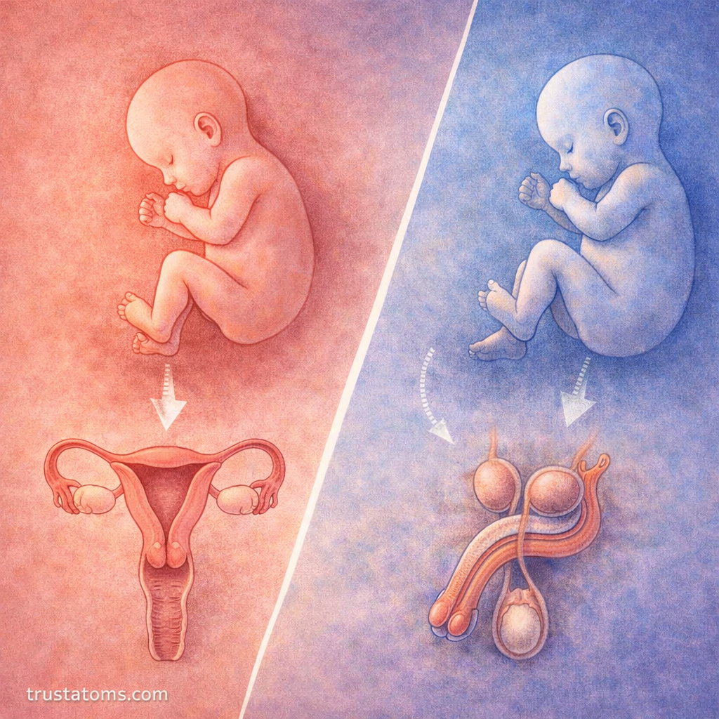
The two embryonic duct systems respond differently to fetal hormones, producing distinct internal reproductive structures.
| Embryonic Structure | Typical Male Development | Typical Female Development |
|---|---|---|
| Undifferentiated gonad | Testis | Ovary |
| Wolffian ducts | Epididymis, vas deferens, seminal vesicles | Mostly regress |
| Müllerian ducts | Regress in response to AMH | Uterine tubes, uterus, cervix, upper vagina |
These structures later contribute to the movement of sperm, eggs, and reproductive secretions. Reproductive Tract Functions and Transport explains their roles after development.
For a closer examination of the testes, epididymis, and vas deferens, see Male Reproductive Anatomy Overview.
The fetal hormonal pathways that shape these structures differ from the endocrine cycles operating after reproductive maturity. Reproductive Hormones and Regulation explains how hormone signaling controls reproductive function.
Later in life, these organs undergo further changes as hormone levels increase during adolescence. Puberty and Reproductive Maturation describes this later developmental stage.
How External Genitalia Develop
External genitalia arise from shared embryonic structures. Their differentiation depends largely on androgen exposure and the ability of developing tissues to respond to those hormones.
- Genital tubercle: Develops into the penis or clitoris.
- Urogenital folds: Contribute to the penile urethra in typical male development or the labia minora in typical female development.
- Labioscrotal swellings: Form the scrotum or labia majora.
DHT signaling promotes masculinization of these tissues. In the absence of substantial androgen effects, they develop along typical female pathways.
Internal and external differentiation are related but partly independent processes. This helps explain why some developmental variations affect one group of structures more than another.
When Sexual Differentiation Occurs
Sexual differentiation begins during the first trimester and continues as reproductive tissues grow and mature. Developmental timing is commonly described using gestational age, measured from the last menstrual period.
| Approximate Gestational Age | Developmental Event |
|---|---|
| Weeks 5–7 | Early gonadal and reproductive duct structures are developing. |
| Weeks 7–9 | Testicular differentiation and hormone production begin in typical XY development. |
| Weeks 9–12 | Hormonal effects increasingly shape internal ducts and external genitalia. |
| After week 12 | Reproductive structures continue growing and becoming more distinct. |
These periods overlap, and individual structures do not develop on an identical schedule. Embryology textbooks sometimes use fertilization age, which is approximately two weeks less than gestational age.
An Example of How Hormone Response Changes Development
Consider complete androgen insensitivity syndrome (CAIS), in which a person typically has XY chromosomes and testes but cells cannot respond normally to androgens because of changes affecting the androgen receptor.
The testes produce AMH, so Müllerian structures generally regress. However, the body’s inability to respond to testosterone and DHT means external genitalia typically develop along female lines, and Wolffian duct development is limited.
This example shows why chromosomes, gonadal development, hormone production, and tissue sensitivity must be considered separately when explaining reproductive anatomy.
Variations in Sex Development
Differences in sex development (DSDs) are congenital variations involving chromosomal, gonadal, or anatomical sex development. They can arise through several biological mechanisms.
- Chromosomal variations: Differences in sex chromosome number or structure can influence gonadal development.
- Gene variations: Changes in genes involved in gonadal differentiation can alter developmental pathways.
- Hormone production differences: Conditions such as congenital adrenal hyperplasia can change fetal androgen exposure.
- Hormone response differences: Changes in androgen receptors or hormone-converting enzymes can affect tissue differentiation.
These variations differ considerably in their anatomy, health implications, and clinical needs. Understanding their biological causes supports accurate diagnosis and individualized medical care.
Frequently Asked Questions
What determines sexual differentiation in an embryo?
Sexual differentiation depends on chromosomes, developmental genes, gonadal formation, fetal hormone production, and tissue responses. These factors work together to shape internal and external reproductive structures.
Does sexual differentiation happen only during puberty?
No. Major reproductive structures differentiate before birth, especially during the first trimester. Puberty later increases reproductive hormone activity and produces additional physical maturation.
Can sexual differentiation vary from typical XX or XY patterns?
Yes. Variations in chromosomes, genes, hormone synthesis, or hormone sensitivity can produce differences in gonadal or reproductive anatomy. These conditions have distinct causes and do not all produce the same developmental outcomes.
Sexual differentiation is a sequence of developmental events in which genes establish gonadal pathways and hormones shape reproductive ducts and external genitalia. Understanding the timing and interaction of these signals explains both typical reproductive anatomy and the biological mechanisms behind variations in sex development.
This article is for general education and is not medical advice. Talk to a qualified health professional about any medical concern.
Sources: The Developing Human: Clinically Oriented Embryology by Moore, Persaud, and Torchia; Langman’s Medical Embryology by Sadler; Williams Textbook of Endocrinology.