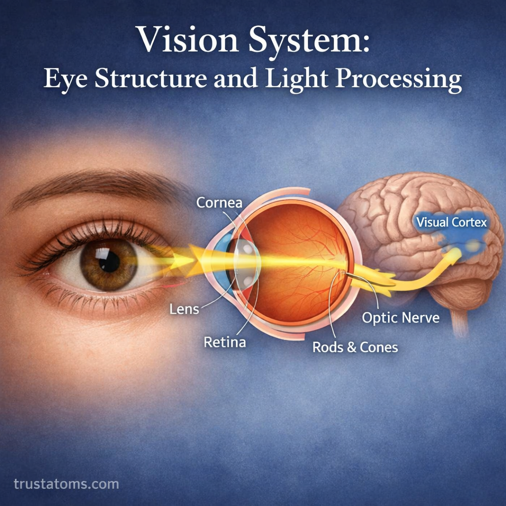
The human vision system works by focusing incoming light onto the retina, where specialized cells convert it into electrical signals that the brain processes into visual information. The cornea, lens, retina, optic nerve, and visual cortex work together to make it possible to recognize colors, shapes, movement, and depth.
Understanding the eye connects human anatomy with the nervous system. From detecting brightness to recognizing familiar faces, vision depends on coordinated eye structures and neural pathways that transmit and process information.
How the Human Vision System Works
Vision begins when light reflected from objects enters the eye. Optical structures focus that light onto the retina, which converts it into signals that travel through the optic nerve to the brain.
The process involves three main components:
- Eyes: Capture and focus light while detecting changes in brightness and color.
- Optic nerves: Carry electrical signals from retinal ganglion cells toward the brain.
- Visual processing regions: Analyze patterns, movement, color, and spatial relationships.
The eye’s photoreceptors are examples of specialized Sensory Receptors that convert environmental stimuli into cellular signals. The Hearing System performs a comparable conversion using sound vibrations rather than light.
External Structures That Protect and Focus the Eye
The outer structures of the eye provide protection, maintain its shape, and begin the process of focusing incoming light.
- Sclera: The tough, white outer layer that protects the eyeball and helps maintain its shape.
- Cornea: The transparent, curved front surface that provides most of the eye’s refractive power.
- Conjunctiva: A thin membrane covering the exposed sclera and lining the inner eyelids, helping protect and lubricate the eye.
The cornea is especially important for clear vision because its curvature bends incoming light toward the retina. Changes in its shape can cause blurred or distorted images.
Internal Eye Anatomy and Its Functions
Inside the eye, several structures regulate light entry, adjust focus, and support the retina.
| Structure | Function |
|---|---|
| Iris | Colored muscular structure that controls pupil size |
| Pupil | Opening that regulates how much light enters |
| Lens | Flexible, transparent structure that fine-tunes focus |
| Aqueous humor | Clear fluid that nourishes nearby tissues and helps maintain eye pressure |
| Vitreous humor | Transparent gel that fills the space behind the lens |
| Retina | Light-sensitive tissue containing photoreceptors and neural circuits |
In bright conditions, the iris constricts the pupil to reduce incoming light. In dim conditions, the pupil dilates to admit more light. The lens also changes shape to keep objects at different distances in focus.
Rods and Cones: How the Retina Detects Light
The retina contains two major types of photoreceptors: rods and cones. Both respond to light, but they are adapted to different visual conditions.
Rods Detect Dim Light
Rods are highly sensitive photoreceptors that support vision in low-light conditions. They contribute strongly to peripheral vision but do not provide color discrimination.
Cones Detect Color and Fine Detail
Cones operate most effectively in brighter light and support detailed color vision. Humans typically have three cone classes with different spectral sensitivities, allowing the visual system to distinguish a wide range of colors.
Cones are especially concentrated in the fovea, a small region near the center of the retina responsible for the sharpest central vision.
Photoreceptors detect light and communicate with other retinal neurons rather than sending signals directly to the brain. Sensory Neurons and Signal Detection explains the broader principles of how sensory information is converted and transmitted.
The Path Light Takes Through the Eye
Before reaching the retina, light passes through several transparent structures that help direct and focus it.
- Cornea: Refracts incoming light, providing most of the eye’s focusing power.
- Aqueous humor: Allows light to pass through the fluid-filled anterior portion of the eye.
- Pupil: Regulates the amount of light entering the eye.
- Lens: Adjusts its curvature to refine focus.
- Vitreous humor: Transmits light through the large posterior chamber.
- Retina: Detects the focused light and begins neural processing.
The eye’s optical system forms an inverted image on the retina. The brain interprets the resulting neural information, allowing us to perceive the environment in its familiar orientation.
How Light Becomes an Electrical Signal
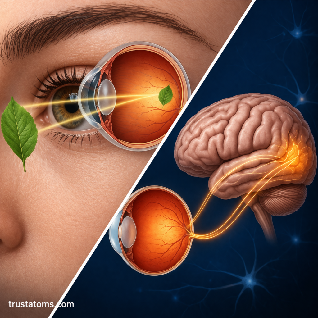
The conversion of light into changes in electrical activity is called phototransduction. It begins when photons are absorbed by light-sensitive pigments inside rods or cones.
Unlike many sensory receptors, photoreceptors become more electrically negative, or hyperpolarized, when stimulated by light. This changes their release of the neurotransmitter glutamate.
Signals are then processed through retinal circuits involving bipolar, horizontal, and amacrine cells. Ganglion cells generate action potentials, and their axons form the optic nerve.
How Visual Signals Reach the Brain
Once retinal ganglion cells generate nerve impulses, visual information travels through a series of connected structures.
- Optic nerve: Carries signals from each eye toward the brain.
- Optic chiasm: Allows fibers from the nasal halves of the retinas to cross to the opposite side.
- Optic tract: Carries information representing the opposite half of the visual field.
- Lateral geniculate nucleus: A region of the thalamus that relays and processes visual signals.
- Visual cortex: Receives signals through optic radiations and begins detailed cortical analysis.
How the Brain Interprets Images
The primary visual cortex is located in the occipital lobe at the back of the brain. It processes basic visual features such as orientation, edges, and spatial patterns.
Information then travels through additional cortical networks that contribute to recognizing objects, detecting movement, and understanding spatial relationships. Cerebral Cortex and Higher Processing examines how cortical regions integrate sensory information.
For example, when catching a moving ball, the visual system estimates its position, speed, and direction. The brain combines this information with motor planning and body-position signals to guide the hands toward the ball.
How Eyes Adjust to Light and Distance
The visual system continually adjusts to changing conditions through several mechanisms.
- Light adaptation: Pupillary responses and changes in photoreceptor sensitivity help vision adjust between bright and dim environments.
- Accommodation: Ciliary muscle activity changes lens shape, increasing its focusing power for nearby objects.
- Binocular depth perception: The brain compares slightly different images from the two eyes to estimate depth.
Common Conditions That Affect Vision
Vision can become impaired when optical structures fail to focus light correctly or when the retina, optic nerve, or brain is affected.
- Myopia: Nearsightedness occurs when distant images focus in front of the retina.
- Hyperopia: Farsightedness occurs when the eye’s optical system would focus images behind the retina without accommodation.
- Astigmatism: Uneven curvature of the cornea or lens causes light to focus differently across orientations.
- Cataracts: Clouding of the lens reduces the clarity of incoming light.
- Glaucoma: Progressive optic nerve damage, often associated with elevated eye pressure, can cause permanent vision loss.
- Age-related macular degeneration: Damage to the macula affects central vision and fine-detail tasks.
Frequently Asked Questions
Which part of the eye converts light into electrical signals?
Photoreceptors called rods and cones in the retina convert light into changes in electrical activity. Retinal neurons process these signals before ganglion cells transmit information through the optic nerve.
Why can humans see colors in daylight but not clearly at night?
Color vision depends mainly on cones, which require relatively bright light. In dim conditions, more sensitive rods dominate visual detection, providing limited color information.
Does the brain or the eye create what we see?
Both contribute. The eye focuses light and begins processing visual signals, while the brain combines and interprets that information to produce visual perception. Neither performs the entire process independently.
Vision depends on a continuous sequence of optical focusing, photoreceptor activity, retinal processing, and interpretation by the brain. Understanding this sequence helps explain why problems affecting the cornea, lens, retina, optic nerve, or visual cortex can produce different kinds of visual impairment.
This article is for general education and is not medical advice. Talk to a qualified health professional about any medical concern.
Sources: Guyton and Hall Textbook of Medical Physiology; Kandel’s Principles of Neural Science; National Eye Institute.