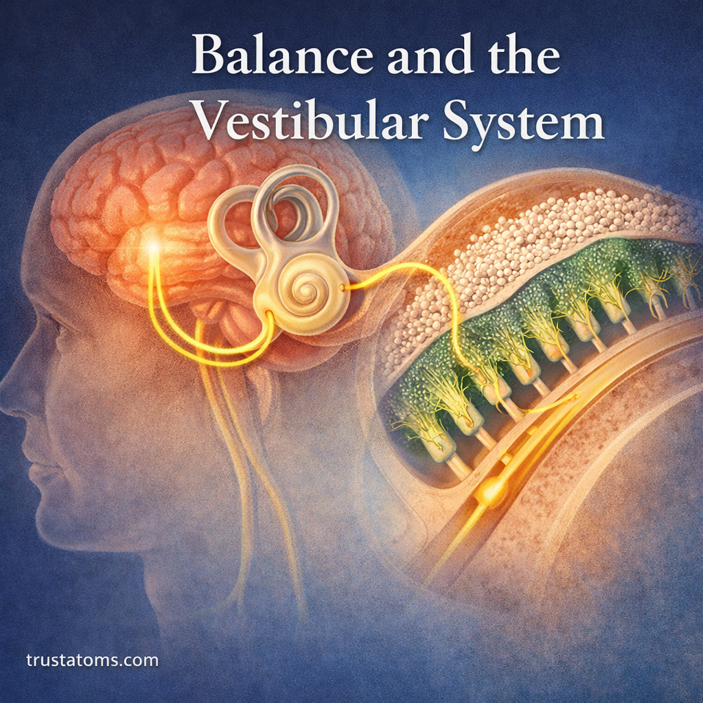
The vestibular system is a sensory system in the inner ear that detects head movement, acceleration, and orientation relative to gravity. It helps maintain balance by sending information to the brain, which combines it with visual and body-position signals to stabilize posture, coordinate movement, and keep vision steady.
Every time you walk, turn your head, or stand on an uneven surface, your brain uses vestibular information to make rapid adjustments. Understanding this system connects human anatomy with neuroscience by explaining how the inner ear and nervous system work together to maintain stability.
What Is the Vestibular System?
The vestibular system consists of specialized sensory organs within the inner ear and neural pathways that carry movement information to the brain. Its receptors detect rotational movement, straight-line acceleration, and head orientation relative to gravity.
The vestibular organs sit beside the cochlea, which detects sound. Although both systems occupy the inner ear, they perform different sensory functions. The Hearing System explains how the cochlea converts sound vibrations into neural signals.
Vestibular information travels through the vestibular branch of the vestibulocochlear nerve, also called cranial nerve VIII. Cranial Nerves and Their Functions describes how this nerve and other cranial nerves connect sensory structures with the brain.
The brainstem and cerebellum process vestibular signals and coordinate reflexes that control eye movements and posture. Cerebellum Function explores how this brain region helps refine movement and balance.
Where the Vestibular Organs Are Located
Each inner ear contains five main vestibular sensory organs: three semicircular ducts and two otolith organs. They occupy fluid-filled structures within the bony labyrinth of the temporal bone.
- Semicircular ducts: Detect angular acceleration associated with head rotation.
- Utricle: Detects linear acceleration and head tilt, particularly in approximately horizontal directions when the head is upright.
- Saccule: Detects linear acceleration and gravity-related forces, particularly along approximately vertical directions when the head is upright.
These structures provide complementary information. Together, they allow the brain to distinguish many patterns of head movement and orientation.
Semicircular Canals: Detecting Head Rotation
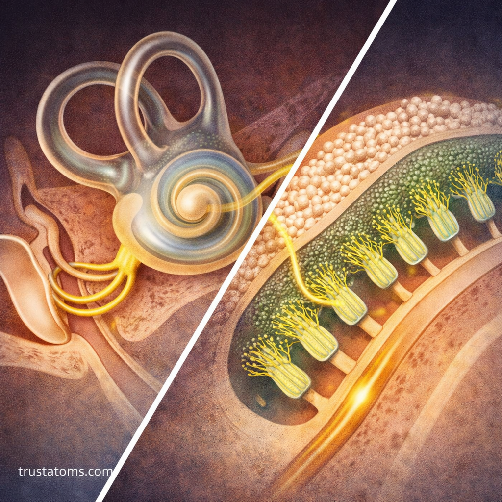
The three semicircular canals are arranged in different planes, allowing them to detect rotations around different axes. Each contains a membranous semicircular duct filled with endolymph.
When the head begins rotating, inertia causes the endolymph to move relative to the duct. This deflects a flexible structure called the cupula, which bends the sensory hair bundles embedded within it.
The resulting changes in nerve signaling inform the brain about angular movement. For example, shaking your head from side to side activates canals sensitive to horizontal rotation, while nodding engages canals oriented to detect other rotational directions.
The canals are especially sensitive to changes in rotational speed. During sustained rotation, their responses gradually adapt, helping explain why spinning sensations can change even while movement continues.
Otolith Organs: Sensing Gravity and Linear Acceleration
The utricle and saccule contain sensory regions called maculae. Each macula has hair cells whose bundles extend into a gelatinous layer containing tiny calcium carbonate crystals called otoconia.
Because this crystal-containing layer has greater mass than the surrounding structures, gravity and linear acceleration can shift it relative to the sensory cells. The resulting bending of hair bundles changes vestibular nerve activity.
These organs respond when you accelerate in a car, ride an elevator, or tilt your head. However, otolith signals alone cannot always distinguish head tilt from linear acceleration. The brain interprets them alongside information from the semicircular canals, vision, and other senses.
Hair Cells Convert Movement Into Nerve Signals
Vestibular hair cells are specialized sensory receptors. Their hair bundles contain stereocilia and a kinocilium, and bending the bundle changes the opening of mechanically sensitive ion channels.
Movement in one direction increases the hair cell’s electrical response and neurotransmitter release, while movement in the opposite direction generally decreases them. Vestibular nerve fibers carry the resulting changes in activity toward the brain.
These signals contribute to rapid adjustments in muscle activity and body position. Balance Control explains how sensory information is translated into postural corrections during standing and movement.
How the Brain Processes Vestibular Information
Vestibular nerve fibers project to vestibular nuclei in the brainstem and directly to parts of the cerebellum. These structures help coordinate eye movements, posture, and the interpretation of head motion.
Vestibular pathways also communicate with spinal motor circuits and brain regions involved in spatial orientation. This distributed processing helps the body respond quickly while maintaining awareness of its position.
Integration With Vision and Body Awareness
Balance depends on combining information from three major sensory sources:
- Vestibular signals: Indicate head rotation, acceleration, and orientation relative to gravity.
- Visual signals: Provide information about surroundings and movement relative to the environment.
- Proprioceptive signals: Describe the position and movement of muscles, joints, and other body structures.
Proprioception and Body Position Sensing explains how receptors in muscles and joints help the brain estimate body position.
When visual information becomes unavailable, such as when standing with your eyes closed, the nervous system relies more heavily on vestibular and somatosensory signals. This is one reason balancing on a soft surface can become more difficult without visual cues.
The Vestibulo-Ocular Reflex Keeps Vision Stable
The vestibulo-ocular reflex (VOR) produces eye movements that compensate for head movements. When the head rotates in one direction, the eyes generally move in the opposite direction to help keep an image stable on the retina.
Consider reading a sign while walking. Your head moves slightly with each step, but vestibular signals help adjust your eye position so the letters remain easier to see.
Without an effective VOR, head movement can cause visual blurring or a sensation that the surroundings are bouncing, known as oscillopsia.
An Everyday Example: Balancing on One Foot
Imagine standing on one foot for 10 seconds. Your body makes small adjustments as its center of mass shifts over a narrower base of support.
Vestibular organs detect head movement and orientation, while proprioceptors report changes in the supporting ankle and leg. Vision provides additional reference points. The brain combines these signals to adjust muscle activity and reduce the chance of falling.
Closing your eyes removes visual information, often making the same task harder. This simple comparison demonstrates why balance depends on several sensory systems rather than the inner ear alone.
Common Vestibular Disorders and Symptoms
Vestibular disorders can interfere with balance, spatial orientation, and stable vision. Different conditions affect different parts of the system.
- Benign paroxysmal positional vertigo (BPPV): Displaced otoconia enter a semicircular canal, causing brief episodes of vertigo triggered by certain head positions.
- Vestibular neuritis: Dysfunction of the vestibular nerve, often associated with inflammation, can cause sudden prolonged vertigo and imbalance.
- Ménière’s disease: An inner ear disorder associated with episodes of vertigo, fluctuating hearing symptoms, tinnitus, and a sensation of ear fullness.
Possible symptoms include spinning sensations, nausea, unsteadiness, and difficulty keeping vision clear during movement. Dizziness has many potential causes, including some unrelated to the vestibular system. Sudden dizziness accompanied by weakness, difficulty speaking, severe headache, or other new neurological symptoms requires urgent medical evaluation.
Frequently Asked Questions
Is the vestibular system responsible for all balance?
No. Vestibular information is one component of balance. Vision, proprioception, muscle strength, and coordinated nervous system responses also contribute to keeping the body upright.
Why can spinning make you feel dizzy afterward?
After rotation stops, fluid movement within the semicircular ducts can continue briefly. This may stimulate hair cells in a way that produces a temporary sensation of continued or opposite rotation.
Can the brain adapt to vestibular problems?
Yes. The nervous system can sometimes compensate for altered vestibular input through adaptation and greater use of other sensory information. Vestibular rehabilitation exercises may help certain patients, depending on the cause of their symptoms.
The vestibular system supplies the brain with continuous information about head motion and orientation, allowing rapid adjustments in eye movements and posture. Its semicircular canals, otolith organs, and sensory hair cells work with visual and proprioceptive pathways to maintain stability. When these signals become disrupted, even ordinary activities such as walking or turning the head can become difficult.
This article is for general education and is not medical advice. Talk to a qualified health professional about any medical concern.
Sources: National Institute on Deafness and Other Communication Disorders, Balance Disorders; Purves and colleagues, Neuroscience, Oxford University Press; Kandel and colleagues, Principles of Neural Science, McGraw Hill.