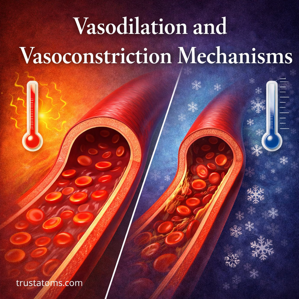
Vasodilation and Vasoconstriction Mechanisms

Illustration comparing vasodilation and vasoconstriction showing widened and narrowed blood vessels with blood flow differences.
Diagram comparing vasodilation and vasoconstriction in blood vessels. trustatoms.com

Blood vessels are not static tubes—they actively adjust their diameter to regulate blood flow, pressure, and temperature. Two key processes control this behavior: vasodilation and vasoconstriction.

These mechanisms are essential for maintaining homeostasis, allowing the body to respond quickly to changes such as exercise, stress, or environmental temperature.

Continue reading “Vasodilation and Vasoconstriction Mechanisms”
Blood Flow Dynamics and Laminar Flow

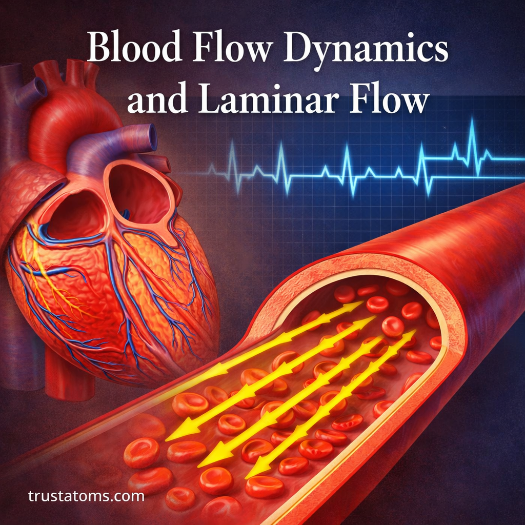
Illustration of blood flow dynamics showing laminar flow in a blood vessel with red blood cells moving in parallel layers and a heart in the background.
Laminar blood flow moves in orderly layers, with blood traveling fastest near the center of a vessel.

Blood flow dynamics describes how blood moves through vessels in response to pressure differences, resistance, and the physical properties of blood. Laminar flow is an orderly pattern of movement in which blood travels in layers, generally moving fastest near the vessel’s center and slowest near its walls.

Understanding these patterns connects human anatomy with cardiovascular physiology. The heart generates pressure, blood vessels regulate resistance, and changing flow conditions determine how oxygen and nutrients reach tissues throughout the body.

Continue reading “Blood Flow Dynamics and Laminar Flow”

Cardiac Conduction System: SA Node to Purkinje Fibers

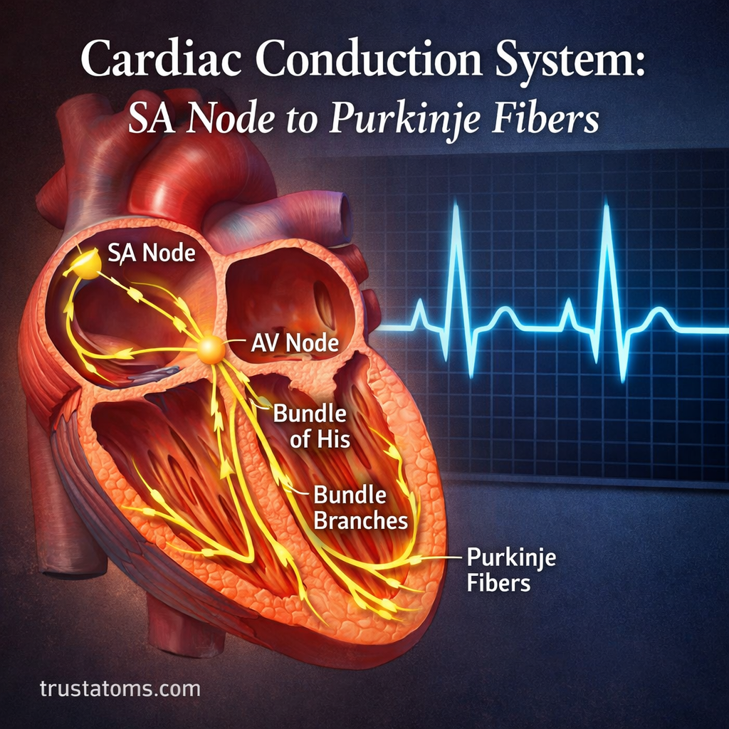
Illustration of the cardiac conduction system showing SA node, AV node, bundle of His, bundle branches, and Purkinje fibers with electrical pathways.
Electrical impulses normally travel from the SA node through the AV node, bundle of His, bundle branches, and Purkinje fibers to coordinate heart contractions.

The cardiac conduction system is a network of specialized heart cells that generates and distributes electrical impulses to coordinate each heartbeat. Signals normally begin in the sinoatrial (SA) node, spread through the atria, pause at the atrioventricular (AV) node, and travel through the bundle of His, bundle branches, and Purkinje fibers to activate the ventricles.

This sequence allows the upper heart chambers to contract before the lower chambers, supporting efficient blood pumping. Understanding the pathway connects electrical signaling with human anatomy and explains how the heart maintains its rhythm without requiring a separate nerve impulse from the brain for every beat.

Continue reading “Cardiac Conduction System: SA Node to Purkinje Fibers”

Electrocardiogram Basics: Electrical Conduction Pathways

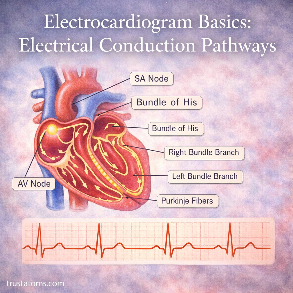
Illustration of the heart’s electrical conduction system showing SA node, AV node, bundle of His, bundle branches, and Purkinje fibers with ECG waveform.
Diagram of the heart’s electrical conduction pathways and ECG waveform showing how signals control heartbeat rhythm.

An electrocardiogram (ECG or EKG) records electrical activity generated by the heart as impulses travel through its conduction system and activate the heart muscle. Its characteristic P waves, QRS complexes, and T waves reveal the timing of atrial and ventricular electrical activity, helping clinicians assess heart rate, rhythm, and conduction abnormalities.

Understanding these signals connects the heart’s internal structures with their functions in human anatomy. An ECG does not directly measure blood flow or the strength of heart contractions, but it provides information about the electrical events that coordinate each heartbeat.

Continue reading “Electrocardiogram Basics: Electrical Conduction Pathways”

Carbon Dioxide Transport in Blood

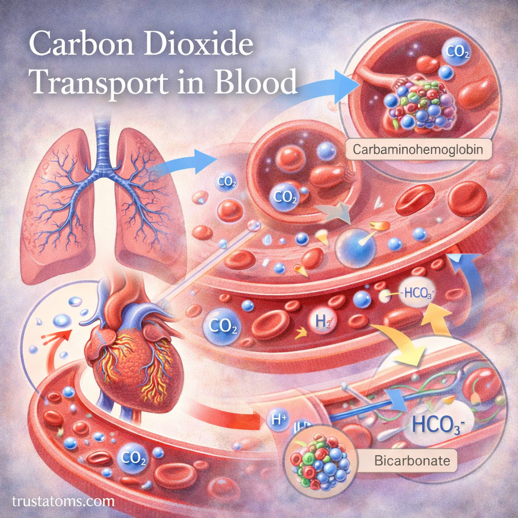
Illustration showing carbon dioxide transport in blood including dissolved CO2, carbaminohemoglobin, and bicarbonate (HCO3-) formation.
Blood transports carbon dioxide mainly as bicarbonate ions, with smaller amounts dissolved directly or attached to proteins such as hemoglobin.

Carbon dioxide (CO₂) travels through the bloodstream in three main forms: bicarbonate ions, CO₂ bound to proteins, and dissolved CO₂. Most is converted into bicarbonate inside red blood cells, allowing blood to carry carbon dioxide from active tissues to the lungs for exhalation.

This transport process also helps regulate blood acidity because carbon dioxide participates in chemical reactions involving hydrogen ions. Understanding these reactions connects respiratory physiology with human anatomy, showing how blood, lungs, and tissues cooperate to maintain stable internal conditions.

Continue reading “Carbon Dioxide Transport in Blood”

Oxygen Delivery and Tissue Demand

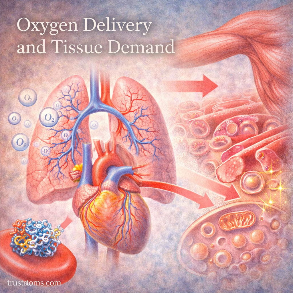
Illustration showing oxygen delivery from the lungs and heart to body tissues and cells where energy is produced.
Diagram of how oxygen moves from the lungs through the bloodstream to tissues to meet cellular energy demands. trustatoms.com.

Oxygen delivery is a fundamental process that allows cells to produce energy and maintain normal function. Every tissue in the body depends on a steady supply of oxygen, and the body continuously adjusts blood flow to match changing demands.

This balance between oxygen delivery and tissue demand is essential for survival, especially during activities like exercise, stress, or illness.

Continue reading “Oxygen Delivery and Tissue Demand”

Baroreceptors and Reflex Control of Pressure

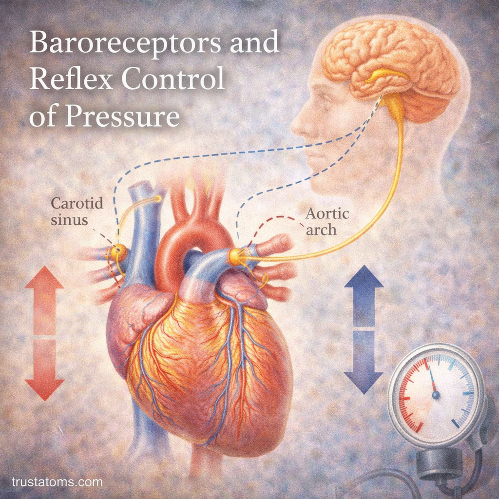
Illustration of baroreceptors in the carotid sinus and aortic arch regulating blood pressure through reflex control involving the heart and brain.
Baroreceptors in the carotid sinus and aortic arch detect arterial stretch and send signals to the brainstem to help regulate blood pressure.

Baroreceptors are specialized sensory nerve endings that detect changes in blood vessel stretch and help regulate blood pressure through a rapid reflex. When arterial pressure rises or falls, they signal the brainstem, which adjusts heart rate, cardiac activity, and blood vessel constriction to stabilize circulation.

This automatic feedback system helps maintain blood flow to organs when you stand, exercise, or experience sudden changes in circulation. Understanding baroreceptors connects human anatomy with cardiovascular physiology by showing how sensory nerves and autonomic pathways work together to control arterial pressure.

Continue reading “Baroreceptors and Reflex Control of Pressure”

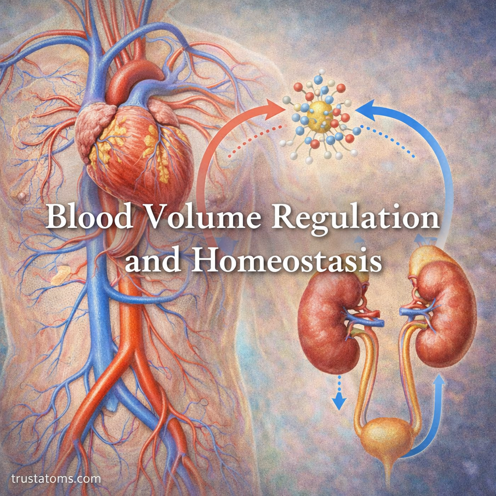
Blood Volume Regulation and Homeostasis

Illustration showing blood volume regulation and homeostasis with the heart, kidneys, and fluid balance processes.
The kidneys, heart, blood vessels, and hormones coordinate fluid retention and excretion to regulate circulating blood volume.

Blood volume regulation is the process by which the body maintains an appropriate amount of circulating blood by adjusting fluid intake, kidney function, and hormonal signals. It supports homeostasis by helping preserve blood pressure, tissue perfusion, and the balance of water and electrolytes.

This process connects human anatomy with the functions of the kidneys, cardiovascular system, and endocrine system. When fluid is lost or retained, these systems respond together to help keep circulation stable.

Continue reading “Blood Volume Regulation and Homeostasis”

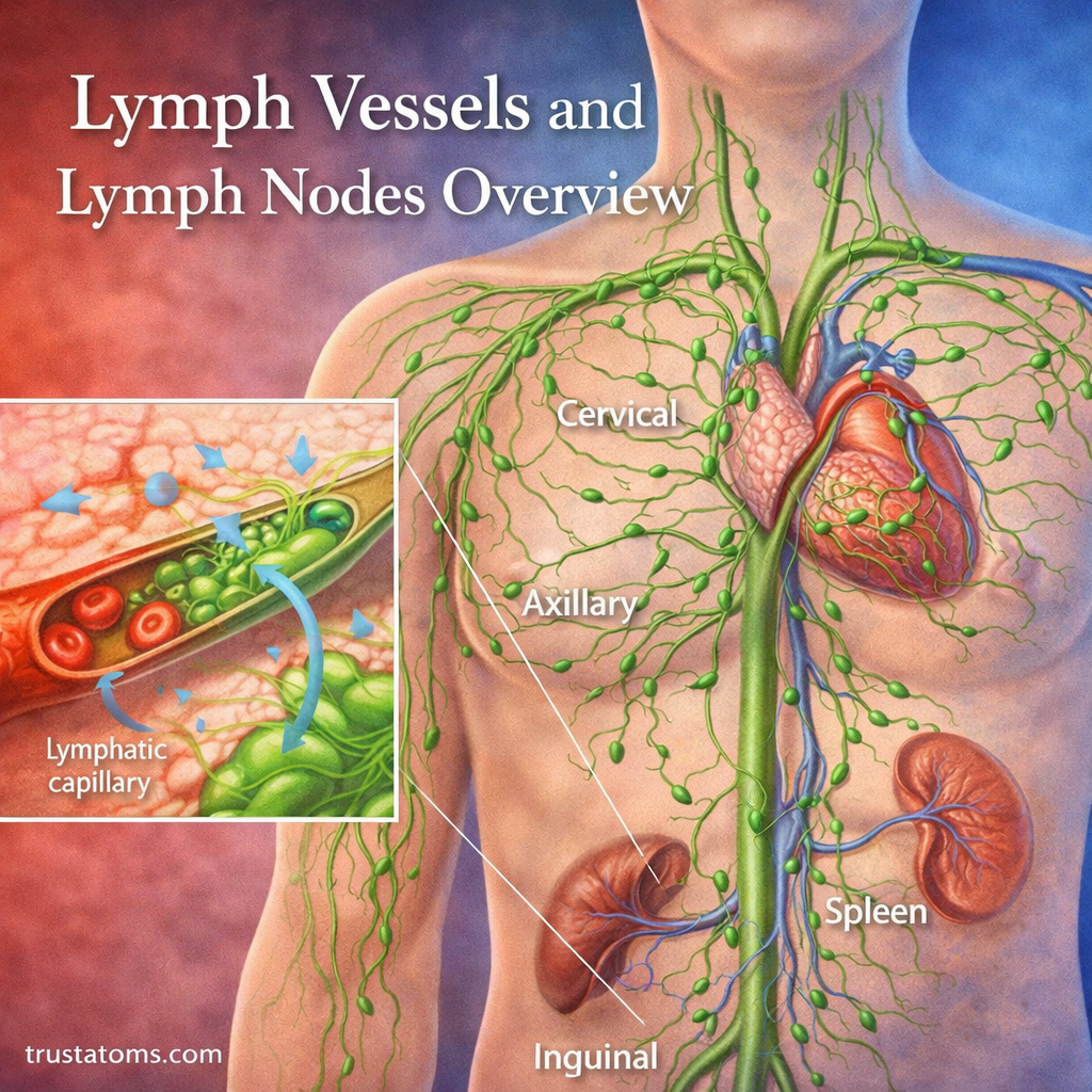
Lymph Vessels and Lymph Nodes Overview

Illustration of lymph vessels and lymph nodes in the human body showing lymph flow, node locations, and capillary interaction.
Overview of lymph vessels and lymph nodes illustrating fluid transport and immune filtration across the body. trustatoms.com.

The lymphatic system is a critical part of human anatomy that supports fluid balance and immune defense. At the core of this system are lymph vessels and lymph nodes—structures that work together to transport lymph and protect the body from harmful invaders.

Understanding how these components function provides insight into how the body maintains internal stability and responds to infection.

Continue reading “Lymph Vessels and Lymph Nodes Overview”

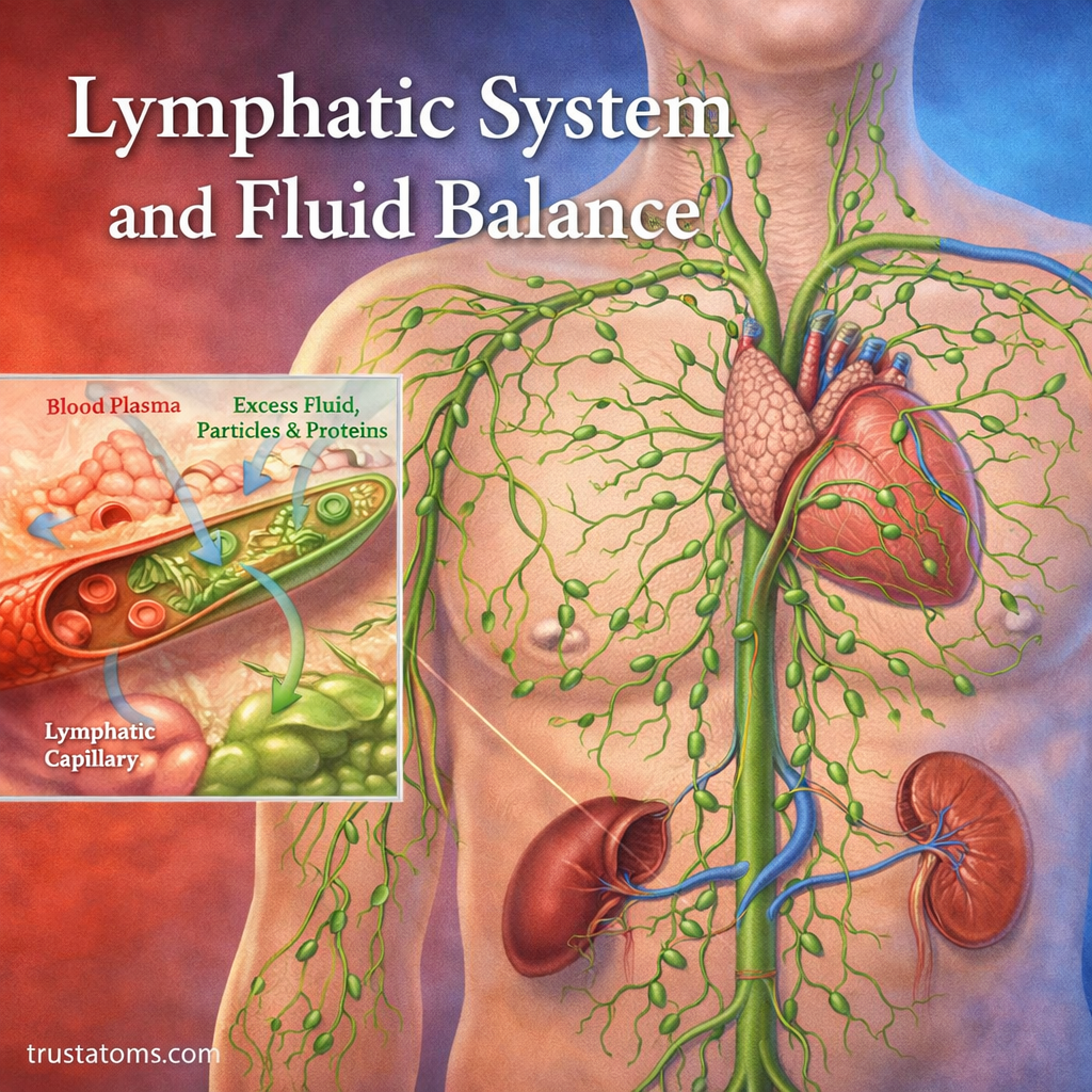
Lymphatic System and Fluid Balance

Illustration of the lymphatic system showing lymph vessels, nodes, and fluid movement from tissues back to the bloodstream.
Overview of the lymphatic system and its role in maintaining fluid balance and immune function. trustatoms.com.

The lymphatic system plays a crucial role in maintaining fluid balance, supporting immune defense, and removing waste from tissues. While often overlooked compared to the circulatory system, it acts as a vital partner in keeping the body stable and functioning efficiently.

Without the lymphatic system, excess fluid would accumulate in tissues, leading to swelling and impaired cellular function.

Continue reading “Lymphatic System and Fluid Balance”