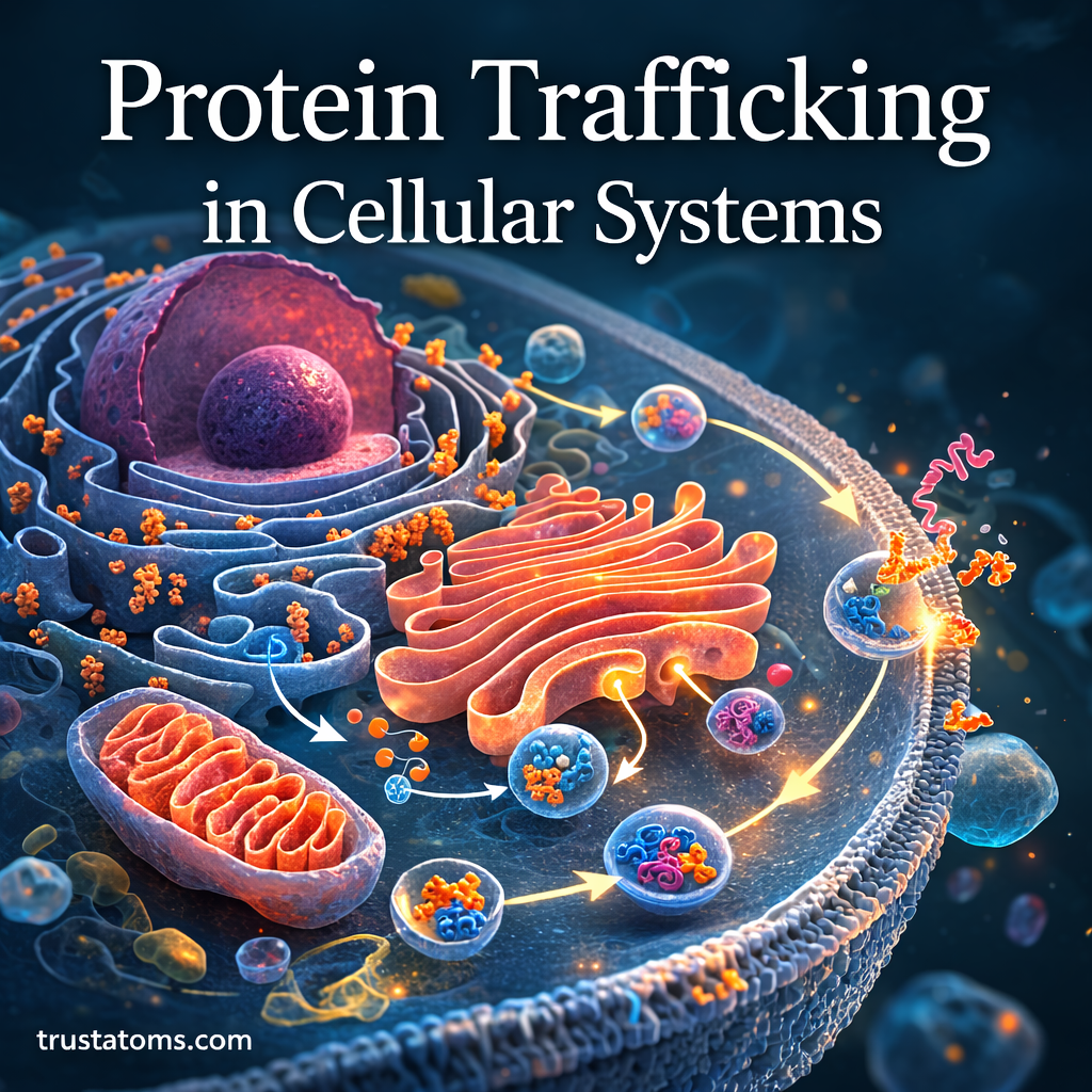
Protein trafficking is the process by which cells move proteins to the locations where they are needed. It includes sorting, transporting, and delivering proteins to organelles, cell membranes, or outside the cell, making it an essential part of biology and cellular organization.
Proteins perform many jobs, including accelerating chemical reactions, transporting substances, and regulating communication between cells. However, a protein must reach the appropriate location to perform its intended function. Cells accomplish this through molecular signals, specialized transport machinery, and networks of membrane-bound compartments.
Some proteins travel through the endoplasmic reticulum and Golgi apparatus, while others enter the nucleus or mitochondria through specialized transport systems. Understanding these different routes helps explain how cells maintain their organization and why transport errors can contribute to disease.
What Is Protein Trafficking?
Protein trafficking describes the movement and sorting of proteins within eukaryotic cells. It begins when proteins are synthesized and continues as they are processed, transported, recycled, or delivered to their functional destinations.
Possible destinations include the cytosol, nucleus, mitochondria, endoplasmic reticulum, Golgi apparatus, lysosomes, plasma membrane, and extracellular environment. Not every protein follows the same route, and some remain near the location where they were produced.
Protein movement supports the cooperation between organelles described in Organelle Coordination in Cellular Metabolism. It also depends on the compartmental organization explored in Cellular Structural Organization.
Where Protein Trafficking Begins: Ribosomes
Proteins are assembled by ribosomes, which translate messenger RNA instructions into chains of amino acids. In eukaryotic cells, ribosomes may be free in the cytosol or attached to the rough endoplasmic reticulum, also called the rough ER.
Proteins Made by Free Ribosomes
Many proteins synthesized by free ribosomes remain in the cytosol. Others are imported into organelles such as the nucleus, mitochondria, or peroxisomes after or during their synthesis.
These proteins may contain targeting information recognized by transport receptors. For example, many mitochondrial proteins carry sequences that help direct them toward mitochondrial import machinery.
Proteins Entering the Rough ER
Proteins destined for secretion, the plasma membrane, or much of the endomembrane system commonly enter the ER during synthesis. A signal recognition particle helps direct the ribosome and growing protein to the ER membrane, where a channel allows the protein to enter the ER lumen or become inserted into the membrane.
Ribosomes themselves are not permanently divided into free and ER-bound types. Their association with the ER depends on the proteins they are producing.
How Signal Sequences Direct Protein Targeting
Cells use targeting signals to identify where many proteins belong. These signals often consist of particular amino acid sequences, although some depend on a protein’s folded structure.
Examples include ER signal peptides, nuclear localization signals, mitochondrial targeting sequences, and peroxisomal targeting signals. Specialized receptors recognize these signals and connect proteins with the appropriate transport machinery.
The recognition and interpretation of these molecular addresses are examined in Protein Targeting, which explains how cells distinguish between different protein destinations.
The Endomembrane System and Protein Transport
The endomembrane system consists of cellular compartments that exchange materials through membrane trafficking. Major components include the ER, Golgi apparatus, endosomes, lysosomes, transport vesicles, and plasma membrane.
Proteins can move between these compartments through vesicular carriers and other transport intermediates. These movements are part of the broader processes described in Intracellular Protein Transport Mechanisms.
Importantly, not all organelles belong to the endomembrane system. Mitochondria and the nuclear interior use distinct protein import pathways rather than receiving most of their proteins through Golgi-derived vesicles.
How Proteins Are Processed in the ER and Golgi
Protein Folding in the Endoplasmic Reticulum
Once proteins enter the ER, many begin folding into functional three-dimensional structures. Molecular chaperones assist this process, while enzymes help form and rearrange disulfide bonds in appropriate proteins.
Some proteins also receive carbohydrate chains through a modification called glycosylation. These modifications can influence protein stability, recognition, and subsequent transport.
Modification and Sorting in the Golgi
Many proteins leaving the ER travel to the Golgi apparatus, where carbohydrate groups and other molecular features may be modified. The Golgi also helps sort cargo toward different destinations, including endosomes, lysosomes, and the cell surface.
Some proteins are retained in or retrieved to earlier compartments rather than continuing toward secretion. This prevents proteins needed in the ER or Golgi from being permanently lost.
How Vesicles Carry Proteins Between Compartments
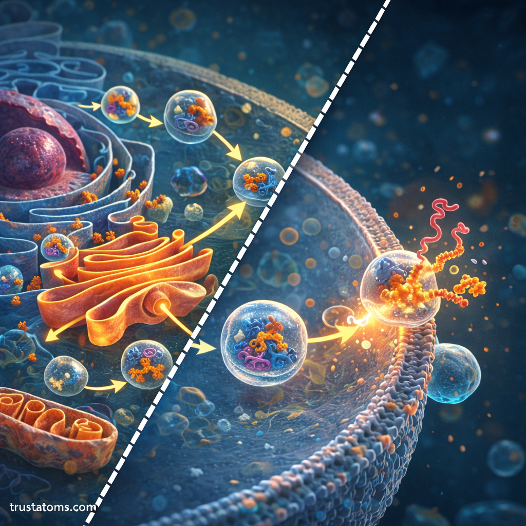
Transport vesicles are small membrane-bound carriers that move selected cargo between cellular compartments. Their formation, movement, recognition, and fusion must be coordinated to deliver proteins accurately.
The basic process involves several stages:
- Cargo selection: Sorting machinery concentrates particular proteins into a developing transport carrier.
- Vesicle formation: Membrane deformation and budding produce a carrier containing selected cargo.
- Transport: The carrier moves toward its destination, sometimes using cytoskeletal tracks and motor proteins.
- Recognition: Regulatory proteins help identify and tether the carrier to the appropriate membrane.
- Membrane fusion: Fusion machinery enables cargo delivery into the destination compartment.
Different vesicle coats support different trafficking routes. COPII commonly participates in ER-to-Golgi transport, while COPI is involved in several retrieval and intra-Golgi pathways. Clathrin-coated carriers participate in selected routes involving the plasma membrane, Golgi, and endosomes.
Rab GTPases help regulate membrane identity and targeting, while SNARE proteins contribute to membrane fusion. Together, these systems support the precision required for vesicle transport throughout the endomembrane system.
Major Protein Trafficking Pathways Compared
Different destinations require different routes. The following table summarizes several common pathways in eukaryotic cells.
| Pathway | Main Transport Route | Example Cargo |
|---|---|---|
| Secretion | ER to Golgi to plasma membrane | Antibodies and secreted enzymes |
| Lysosomal delivery | ER to Golgi to endosomes and lysosomes | Digestive enzymes |
| Nuclear import | Cytosol through nuclear pores | Transcription factors |
| Mitochondrial import | Cytosol through mitochondrial import machinery | Metabolic enzymes |
| Membrane recycling | Plasma membrane to endosomes and back | Cell-surface receptors |
These routes may intersect with recycling and degradation pathways. A protein’s location can also change in response to cellular signals rather than remaining permanently fixed.
Real-World Example: How Cells Secrete Insulin
Insulin secretion provides a concrete example of protein trafficking. Specialized beta cells in the pancreas produce insulin and release it into the bloodstream when stimulated by increased blood glucose.
The pathway begins when ribosomes synthesize preproinsulin and direct it into the ER. Its signal peptide is removed, producing proinsulin, which folds and forms disulfide bonds. Proinsulin then moves through the Golgi into secretory granules.
Within the secretory pathway, proinsulin is processed into insulin and C-peptide. When glucose stimulates the beta cell, cellular signaling increases calcium-dependent fusion of insulin-containing granules with the plasma membrane.
Insulin is then released outside the cell through exocytosis. This example shows how synthesis, folding, processing, storage, and regulated membrane fusion work together to deliver a functional protein at the appropriate time.
How Cells Control Protein Quality and Recycling
Not every newly synthesized protein folds or travels correctly. Cells use quality-control systems to recognize defective proteins and prevent their accumulation.
In the ER, certain misfolded proteins are retained and directed toward ER-associated degradation, which commonly involves the proteasome. Other damaged proteins or cellular components may be broken down through lysosomal pathways.
Cells also recycle many transport receptors and membrane proteins. Recycling reduces unnecessary protein replacement and allows cells to adjust the composition of their membranes in response to changing conditions.
What Happens When Protein Trafficking Fails?
Disrupted trafficking can prevent proteins from reaching their destinations, interfere with cellular communication, or cause material to accumulate in inappropriate compartments.
In cystic fibrosis, the common F508del variant of the CFTR protein causes folding and processing defects that reduce delivery of functional CFTR to the cell surface. Some medicines improve the processing or activity of particular CFTR variants.
Other disorders involve defective lysosomal enzyme delivery, abnormal membrane recycling, or disrupted intracellular transport. Researchers also investigate how trafficking abnormalities contribute to certain neurological diseases, although their mechanisms differ substantially.
How Scientists Observe Protein Movement
Researchers study trafficking using fluorescent protein tagging, live-cell microscopy, biochemical fractionation, and genetic experiments. These methods can reveal where proteins accumulate, how quickly they move, and which transport components are required.
Electron microscopy provides detailed views of cellular structures, while molecular experiments help identify the machinery involved in cargo sorting and membrane fusion. Combining several techniques gives a more reliable picture than relying on protein location alone.
Frequently Asked Questions
What is the difference between protein trafficking and protein targeting?
Protein targeting concerns how cells identify a protein’s intended destination. Protein trafficking includes the broader movement, sorting, delivery, and recycling processes that allow proteins to reach or move between cellular locations.
Do all proteins pass through the Golgi apparatus?
No. Many proteins remain in the cytosol or enter the nucleus, mitochondria, and other organelles without passing through the Golgi. The Golgi primarily processes and sorts proteins traveling through the secretory and endomembrane pathways.
What is the difference between secretion and exocytosis?
Secretion refers broadly to the release of substances from cells. Exocytosis is a specific mechanism in which a membrane-bound carrier fuses with the plasma membrane, releasing its contents outside the cell or adding membrane components to the cell surface.
Protein trafficking allows cells to coordinate protein production with delivery, recycling, and removal. Through a coordinated system involving signal sequences, organelle networks, vesicle transport, and quality control mechanisms, cells maintain precise control over protein distribution. Understanding these pathways helps explain normal cellular activity and the consequences of transport defects.
Sources: Alberts et al., Molecular Biology of the Cell; Lodish et al., Molecular Cell Biology; National Institute of General Medical Sciences.