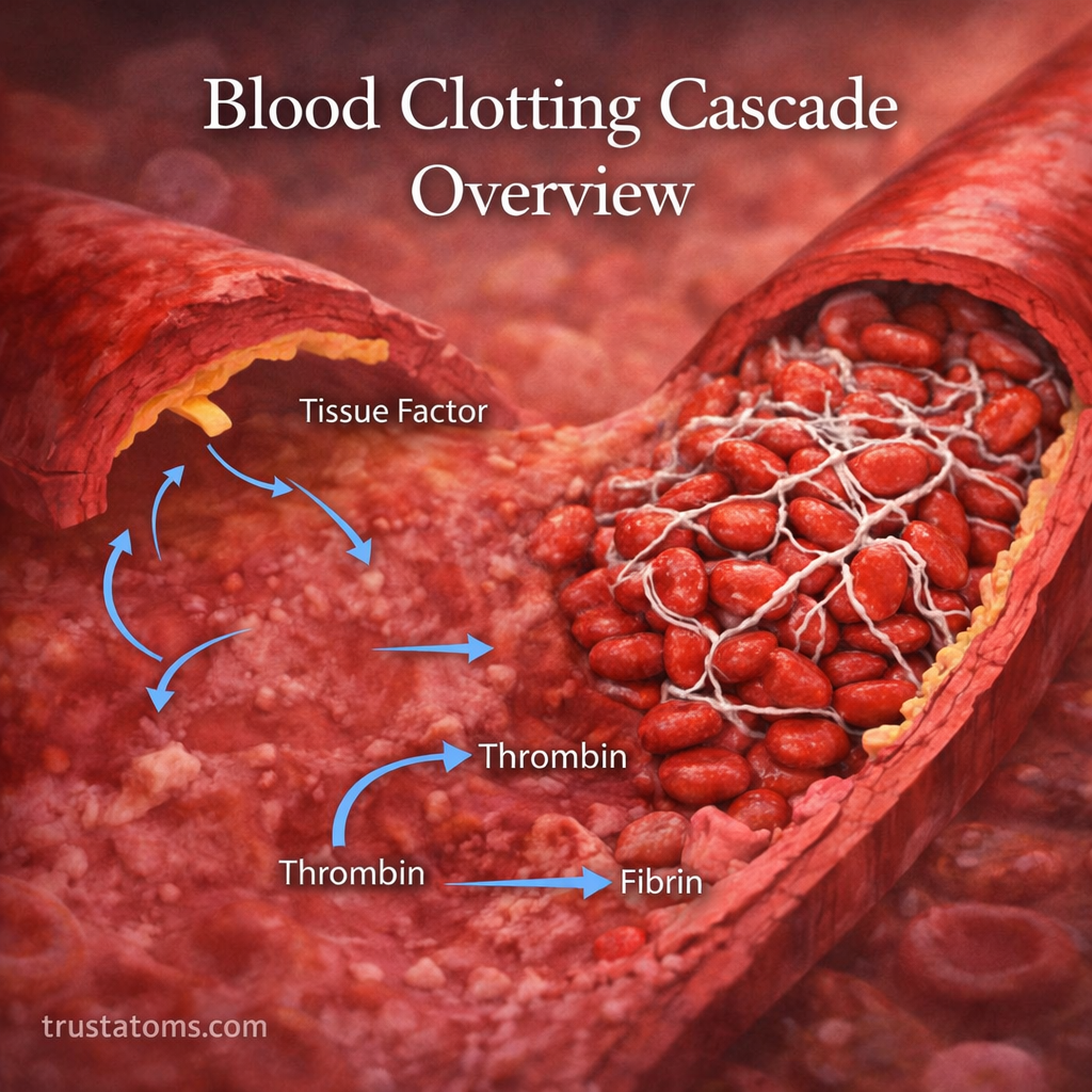
The blood clotting cascade is a series of chemical reactions that produces fibrin, a protein that strengthens blood clots and helps stop bleeding after an injury. It works by activating clotting factors in a coordinated sequence, ultimately converting a temporary platelet plug into a more stable clot.
As part of human anatomy and physiology, this process protects damaged blood vessels while allowing normal circulation elsewhere. When a vessel is injured, the body responds through a complex chain reaction known as the blood clotting cascade. While platelets form the initial plug, the clotting cascade strengthens and stabilizes it, helping control bleeding during tissue repair.
What Is the Blood Clotting Cascade?
The clotting cascade is part of hemostasis, the body’s process for stopping blood loss. It involves clotting factors, most of which are proteins circulating in blood plasma. Many remain inactive until an injury triggers their activation.
Each activated factor helps drive the next reaction, amplifying the response. The result is fibrin, which forms a network around the platelet plug. For additional context, blood composition explains how plasma, platelets, and blood cells contribute to circulation.
Clotting is also an early stage of tissue repair. The wound healing phases describe what happens as damaged skin recovers. Meanwhile, immune defenses respond to injury and potential infection through processes such as those explained in the complement system overview.
The Three Pathways of Blood Clotting
Traditional diagrams divide the cascade into intrinsic, extrinsic, and common pathways. This model helps explain laboratory tests and clotting-factor interactions, although the pathways overlap during clot formation inside the body.
Intrinsic Pathway: Amplifying the Response
The intrinsic pathway is also called the contact activation pathway. In laboratory testing, contact with certain negatively charged surfaces activates Factor XII, followed by reactions involving Factors XI, IX, and VIII.
Within the body, Factors IX and VIII help amplify clotting after tissue-factor signaling begins. Factor XII is useful for understanding laboratory clotting tests but is not essential for normal injury-related hemostasis.
Extrinsic Pathway: Starting Clot Formation
The extrinsic pathway begins when damaged tissue exposes tissue factor, also called Factor III, to circulating blood. Tissue factor binds Factor VII and promotes its activation, initiating reactions that activate Factor X and Factor IX.
This tissue-factor pathway provides the main physiological trigger for coagulation after blood vessel injury.
Common Pathway: Producing a Fibrin Clot
The common pathway begins with activated Factor X (Xa). Working with activated Factor V, calcium, and phospholipid surfaces, Factor Xa helps convert prothrombin (Factor II) into thrombin.
Thrombin converts soluble fibrinogen (Factor I) into fibrin strands. Activated Factor XIII then cross-links these strands, reinforcing the clot so it can better withstand blood flow.
Blood Clotting Cascade: Step-by-Step Example
Consider a small cut on your finger while preparing food. The injured blood vessel constricts, platelets adhere to the damaged area, and clotting proteins begin forming a fibrin network. The process follows these overlapping stages:
- Vessel injury: Damage exposes collagen and tissue factor, initiating hemostasis.
- Platelet plug: Platelets attach, activate, and gather at the injury.
- Factor activation: Tissue factor and Factor VII initiate coagulation, while additional factors amplify it.
- Thrombin generation: Activated Factor X supports the conversion of prothrombin into thrombin.
- Fibrin formation: Thrombin converts fibrinogen into strands that surround the platelet plug.
- Clot stabilization: Factor XIIIa cross-links fibrin, strengthening the clot while tissue repair begins.
The visible scab that may develop afterward is not the clotting cascade itself. It is a protective covering associated with the healing wound.
Important Clotting Factors and Their Functions
| Factor | Main function |
|---|---|
| I (Fibrinogen) | Converted into fibrin |
| II (Prothrombin) | Precursor of thrombin |
| III (Tissue factor) | Initiates tissue-factor signaling |
| VII | Works with tissue factor |
| VIII and IX | Help activate Factor X |
| X | Supports thrombin generation |
| XIII | Cross-links and stabilizes fibrin |
Most clotting factors are produced in the liver. Vitamin K is needed for the normal production of functional Factors II, VII, IX, and X, along with the anticoagulant proteins C and S. Not every clotting factor depends on vitamin K.
How Thrombin Strengthens Blood Clots
Thrombin is a central enzyme in coagulation. Besides producing fibrin, it activates platelets and several clotting factors, including V, VIII, XI, and XIII. These feedback reactions increase fibrin production near the injury.
Thrombin can also participate in anticoagulant regulation when it binds thrombomodulin on healthy endothelial cells, helping activate protein C. Its effects therefore depend on where and how it is generated.
How the Body Prevents Excessive Clotting
Clotting must remain concentrated near damaged tissue. Several protective mechanisms limit the reaction:
- Antithrombin: Inhibits thrombin and other activated clotting factors.
- Protein C and protein S: Help inactivate Factors Va and VIIIa.
- Healthy endothelium: Produces signals that discourage unnecessary platelet activation and clot formation.
- Fibrinolysis: Uses plasmin to break down fibrin as the clot is no longer needed.
These systems help prevent a localized repair response from spreading into an unnecessary blockage.
Bleeding Disorders and Abnormal Blood Clots
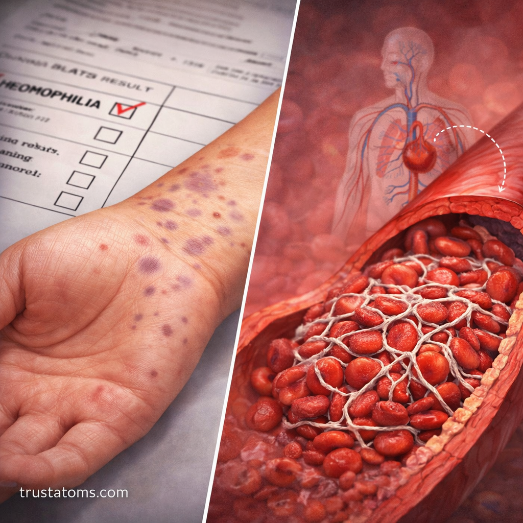
Problems with coagulation can cause either excessive bleeding or unwanted clot formation. Hemophilia A involves reduced Factor VIII activity, while hemophilia B involves Factor IX deficiency. These conditions may cause prolonged bleeding, including bleeding into joints.
Excessive clot formation can cause deep vein thrombosis (DVT), often in a leg. If part of a venous clot travels to the lungs, it may cause a pulmonary embolism. Arterial clots can also contribute to certain heart attacks and ischemic strokes.
Clinicians may investigate coagulation problems using tests such as prothrombin time (PT) and activated partial thromboplastin time (aPTT). These measure different parts of the laboratory clotting pathways and do not independently identify every clotting disorder.
Frequently Asked Questions
What is the difference between the intrinsic and extrinsic pathways?
The intrinsic pathway describes contact-related reactions and amplification involving Factors XI, IX, and VIII. The extrinsic pathway begins with tissue factor and Factor VII and normally initiates coagulation after injury. Both contribute to thrombin generation.
Which clotting factor produces fibrin?
Thrombin, the activated form of Factor II, converts fibrinogen (Factor I) into fibrin. Factor XIIIa then cross-links the fibrin strands to strengthen the clot.
What happens when blood clotting is too slow or too fast?
Insufficient clotting can lead to prolonged or spontaneous bleeding. Excessive clotting may obstruct blood vessels and damage organs. The underlying causes differ and require medical evaluation.
The blood clotting cascade connects vessel injury, platelet activity, and clotting-factor reactions to produce a reinforced fibrin clot. Understanding the tissue-factor trigger, thrombin generation, and fibrin stabilization helps explain both normal wound repair and disorders involving abnormal bleeding or thrombosis.
This article is for general education and is not medical advice. Talk to a qualified health professional about any medical concern.
Sources: Guyton and Hall Textbook of Medical Physiology; Hoffbrand’s Essential Haematology; Merck Manual Professional Edition.