Breathing Control Centers in the Brainstem

Illustration of the brainstem showing the medulla oblongata and pons controlling breathing and sending signals to the diaphragm.
The brainstem regulates breathing automatically through the medulla and pons, controlling respiratory rhythm and depth. trustatoms.com.

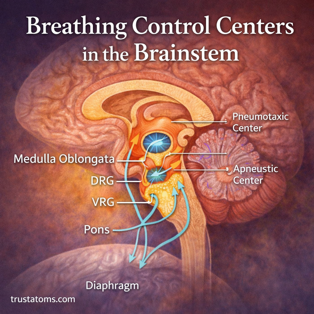
Breathing is something most people rarely think about, yet it happens continuously—day and night—without conscious effort. This automatic process is controlled by specialized regions in the brain known as the breathing control centers, located in the brainstem.

These centers regulate the rhythm, depth, and rate of breathing, ensuring that the body receives enough oxygen and removes carbon dioxide efficiently.

Continue reading “Breathing Control Centers in the Brainstem”

Nasal Filtration and Air Conditioning

Illustration of the nasal cavity showing how the nose filters particles with mucus and hairs while warming and humidifying incoming air.
The nose filters, warms, and humidifies incoming air before it reaches the lungs, supporting respiratory health. trustatoms.com.

Every breath you take passes through a highly efficient system designed to protect and prepare air before it reaches your lungs. The nose does far more than just allow airflow—it acts as a natural filter, humidifier, and temperature regulator.

This process, known as nasal filtration and air conditioning, is essential for maintaining respiratory health and preventing damage to sensitive lung tissues.

Continue reading “Nasal Filtration and Air Conditioning”

Cilia and Mucus in Airway Defense

Illustration of cilia moving mucus with trapped particles in the airway to protect the lungs from dust, bacteria, and viruses.
Cilia and mucus work together in the airway to trap and remove harmful particles, helping protect the lungs. trustatoms.com.

The human respiratory system is constantly exposed to airborne particles such as dust, bacteria, viruses, and pollutants. Despite this, the lungs remain remarkably protected. One of the most effective defense systems responsible for this protection is the coordinated action of cilia and mucus within the airways.

This built-in cleaning mechanism—often called the mucociliary escalator—plays a critical role in keeping the respiratory tract clear and functional.

Continue reading “Cilia and Mucus in Airway Defense”

Pleura and Pleural Pressure

Illustration of pleura and pleural pressure showing lung expansion within the chest cavity and pleural space.
Diagram of the pleural layers and pressure changes that keep the lungs expanded during breathing. trustatoms.com.

The lungs do not function in isolation—they rely on a thin, fluid-filled membrane system called the pleura to move smoothly within the chest cavity. Along with this structure, pleural pressure plays a crucial role in keeping the lungs expanded and enabling efficient breathing.

Understanding the pleura and pleural pressure helps explain how the lungs stay inflated and what happens when this system is disrupted.

Continue reading “Pleura and Pleural Pressure”

Respiratory Muscles Beyond the Diaphragm

Illustration of respiratory muscles including intercostals, neck muscles, and abdominal muscles supporting breathing beyond the diaphragm.
Diagram showing accessory respiratory muscles that assist breathing alongside the diaphragm. trustatoms.com.

When most people think about breathing, they think of the diaphragm—the primary muscle responsible for inhalation. However, breathing also relies on a group of additional muscles that assist in expanding and compressing the lungs, especially during physical activity or respiratory stress.

These supporting muscles, often called accessory respiratory muscles, play a crucial role in maintaining efficient airflow when the body’s demand for oxygen increases.

Continue reading “Respiratory Muscles Beyond the Diaphragm”

Ventilation–Perfusion Matching Principles

Illustration showing ventilation and perfusion matching in alveoli with comparison of normal and impaired airflow and blood flow.
Diagram of ventilation–perfusion matching showing how balanced airflow and blood flow optimize gas exchange in the lungs. trustatoms.com.

Ventilation–perfusion (V/Q) matching is a key principle of respiratory physiology that ensures oxygen enters the blood efficiently and carbon dioxide is removed effectively. It refers to the balance between airflow reaching the alveoli (ventilation) and blood flow reaching the lungs (perfusion).

When ventilation and perfusion are properly matched, gas exchange is optimized. When they are not, breathing becomes less efficient—even if the lungs themselves appear structurally normal.

Continue reading “Ventilation–Perfusion Matching Principles”

Partial Pressure and Gas Diffusion

Illustration showing partial pressure and gas diffusion between alveoli and capillaries with oxygen and carbon dioxide movement.
Diagram of gas exchange in the lungs illustrating how partial pressure drives oxygen into the blood and carbon dioxide out. trustatoms.com.

Gas exchange in the lungs depends on a fundamental principle of physics: gases move from areas of higher pressure to areas of lower pressure. In the respiratory system, this process is driven by differences in partial pressure, allowing oxygen to enter the blood and carbon dioxide to leave it.

Understanding partial pressure and gas diffusion is essential for explaining how breathing supports life at a cellular level.

Continue reading “Partial Pressure and Gas Diffusion”

Surfactant Function in Alveolar Stability

Illustration of surfactant function in alveoli showing reduced surface tension and stabilized air sacs in the lungs.
Close-up of alveoli demonstrating how surfactant reduces surface tension to keep air sacs open and stable. trustatoms.com.

Surfactant is a specialized substance in the lungs that plays a critical role in keeping the alveoli stable and functional. Without it, breathing would require significantly more effort, and the tiny air sacs responsible for gas exchange could collapse.

Understanding how surfactant works provides key insight into respiratory efficiency, lung development, and conditions that affect breathing.

Continue reading “Surfactant Function in Alveolar Stability”

Alveolar Structure and Surface Area

Illustration of alveoli structure and surface area showing air sacs and surrounding capillaries in the lungs.
Detailed view of alveoli highlighting their structure and large surface area for efficient gas exchange. trustatoms.com.

Alveoli are tiny air sacs in the lungs where oxygen and carbon dioxide are exchanged between the air and the bloodstream. Although each alveolus is microscopic, their combined structure creates a massive surface area that is essential for efficient respiration.

Understanding alveolar structure and surface area helps explain how the lungs meet the body’s oxygen demands—and why even small damage to this system can have major consequences.

Continue reading “Alveolar Structure and Surface Area”

Bronchioles and Airway Resistance

Illustration showing bronchioles and airway resistance with normal and constricted airways in the lungs.
Simple diagram of bronchioles demonstrating how airway narrowing increases resistance in the lungs. trustatoms.com.

Bronchioles are the smallest airways in the respiratory system, playing a critical role in regulating airflow to the lungs. While they are tiny compared to the trachea and bronchi, their structure and function have a major impact on airway resistance and overall breathing efficiency.

Understanding how bronchioles influence airflow helps explain normal respiration, exercise performance, and common respiratory conditions such as asthma.

Continue reading “Bronchioles and Airway Resistance”