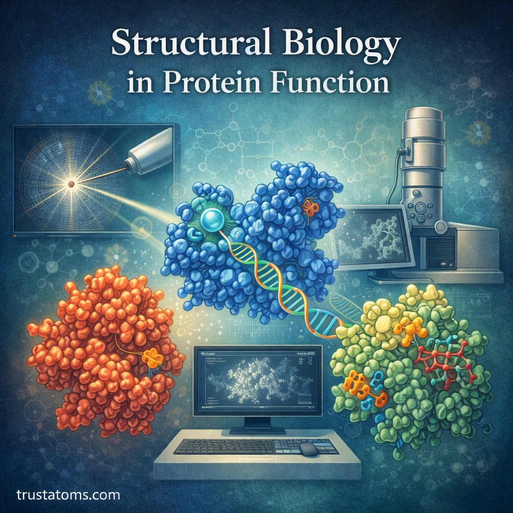
Structural biology explains protein function by revealing how a protein’s three-dimensional shape determines its interactions and biological activity. Scientists use experimental and computational methods to examine molecular structures, identify functional regions, and understand how structural changes affect proteins.
In biological science, understanding molecular structure helps explain processes ranging from enzyme activity to cellular communication. A protein’s amino acid sequence provides the instructions for its structure, but folding, molecular interactions, and surrounding conditions influence how it performs its function.
This article explores the principles of structural biology, the relationship between protein structure and function, and the technologies scientists use to study these molecules.
What Is Structural Biology?
Structural biology is the study of the three-dimensional organization of biological molecules, including proteins, DNA, RNA, and large molecular complexes.
Researchers investigate how atoms are arranged within molecules and how those arrangements influence chemical reactions, molecular recognition, and cellular processes.
Structural biology overlaps with Biophysical Chemistry, which examines the physical and chemical principles governing molecular behavior inside cells.
It also supports the study of Biomolecular Interactions by revealing the surfaces and molecular features involved when proteins, nucleic acids, and other molecules bind together.
Structural information helps researchers explain not only what a biological molecule does, but also how its physical organization makes that activity possible.
Why Protein Structure Matters
Proteins are essential molecules that carry out most of the work inside living cells. They catalyze chemical reactions, transport substances, transmit signals, and provide structural support.
Proteins consist of amino acid chains that fold into specific three-dimensional arrangements. Their shapes influence which molecules they recognize, how efficiently they function, and how they interact with other cellular components.
The process described in Protein Folding in Molecular Systems explains how amino acid sequences and molecular interactions contribute to the formation of functional protein structures.
Protein shape also determines how molecules assemble into larger systems. Research on Protein–Protein Interactions examines how complementary binding surfaces allow proteins to cooperate in cellular pathways.
Even a small structural change can alter a protein’s activity. However, the effects depend on where the change occurs and whether it disrupts folding, stability, or a functional region.
The Four Levels of Protein Structure
Scientists describe protein organization using four structural levels. These levels provide a framework for understanding how amino acid sequences form functional molecules.
Primary Structure
Primary structure is the linear sequence of amino acids connected by peptide bonds.
The sequence influences how the protein folds and which chemical interactions can form. Changing an amino acid may affect the final structure, although some substitutions have little effect.
Secondary Structure
Secondary structure describes local arrangements of the protein backbone, particularly alpha helices and beta sheets.
These structures are stabilized mainly by hydrogen bonds between backbone groups. Loops and turns connect different structural regions.
Tertiary Structure
Tertiary structure describes the overall three-dimensional arrangement of a single polypeptide chain.
It is influenced by hydrophobic interactions, hydrogen bonds, ionic interactions, and sometimes disulfide bonds. These interactions help position functional regions such as enzyme active sites and molecular binding pockets.
Quaternary Structure
Quaternary structure describes how multiple polypeptide chains assemble into a functional protein complex.
Hemoglobin provides a familiar example. Adult hemoglobin A contains four protein subunits: two alpha chains and two beta chains. Their interactions allow the molecule to bind and release oxygen cooperatively.
Not every protein has quaternary structure. Proteins consisting of a single polypeptide chain do not require multiple subunits to function.
How Protein Shape Determines Biological Function
Enzyme Active Sites
Enzymes contain active sites where substrates bind and chemical reactions occur. The arrangement of amino acid side chains helps position substrates and stabilize reaction transition states.
For example, digestive enzymes recognize particular types of molecules. Their active-site structures help determine which chemical bonds they can break and how efficiently the reactions proceed.
Molecular Recognition
Proteins recognize other molecules through complementary shapes and chemical properties, including charge distribution and hydrogen-bonding patterns.
Antibodies recognize particular molecular features on antigens, while receptors bind signaling molecules that influence cellular activity.
Binding often involves small structural adjustments rather than two completely rigid surfaces fitting together.
Protein Stability and Flexibility
Proteins must maintain suitable structures under the conditions in which they function. Changes in temperature, acidity, or chemical environment can alter their stability.
At the same time, flexibility is necessary for many proteins. Enzymes, transport proteins, and molecular motors often change shape during their normal activities.
Techniques Used to Determine Protein Structures

Structural biologists use several techniques to investigate molecular organization. Each method provides different types of information and has practical limitations.
X-ray Crystallography
X-ray crystallography determines molecular structures by analyzing how X-rays diffract through an ordered crystal.
Researchers prepare protein crystals, collect diffraction measurements, and use computational methods to construct an electron-density map and an atomic model.
This method can reveal detailed structural features, but obtaining suitable crystals is often difficult, particularly for flexible or membrane-associated proteins.
Nuclear Magnetic Resonance Spectroscopy
Nuclear magnetic resonance (NMR) spectroscopy examines signals from atomic nuclei in a magnetic field.
Researchers use these signals to investigate molecular structure, interactions, and movement. Solution NMR is especially useful for studying many smaller proteins without requiring crystallization.
Large molecular size and overlapping signals can make some proteins challenging to analyze using conventional solution NMR methods.
Cryo-Electron Microscopy
Cryo-electron microscopy, or cryo-EM, examines biological samples rapidly frozen in a thin layer of vitreous ice.
Electron images of many individual particles are computationally combined to reconstruct three-dimensional structures.
Single-particle cryo-EM is particularly useful for studying large protein complexes and molecular assemblies that are difficult to crystallize.
Computational Structure Prediction
Computational methods can predict protein structures from amino acid sequences. Modern artificial intelligence systems have substantially improved the accuracy of many protein structure predictions.
Predicted structures are useful for developing hypotheses and planning experiments, but they do not automatically reveal every functional state, molecular interaction, or structural movement.
How Structural Biology Supports Drug Discovery
Many medicines work by interacting with specific proteins. Structural information can help researchers identify binding pockets and understand how candidate drugs interact with their targets.
In structure-based drug design, scientists may examine a target protein’s three-dimensional arrangement and design or select molecules that fit a particular binding region.
Researchers then test whether those molecules bind effectively, influence protein activity, and demonstrate suitable biological properties.
A promising structural fit does not guarantee a successful medicine. Drug development also requires evaluating toxicity, stability, delivery, and effects throughout the body.
Structural Biology in Disease Research
Structural studies help scientists investigate how genetic changes and abnormal protein behavior contribute to disease.
For example, sickle cell disease is associated with a substitution of one amino acid in the beta-globin chain of hemoglobin. This change creates a molecular interaction that promotes abnormal hemoglobin assembly under low-oxygen conditions.
Structural research can also reveal how disease-associated mutations affect enzyme activity, receptor signaling, or protein stability.
In neurodegenerative disease research, scientists investigate the structures of misfolded proteins and protein aggregates to better understand their formation and behavior.
Protein Engineering and Biotechnology Applications
Protein engineering involves modifying amino acid sequences to change protein properties. Structural information helps researchers identify regions that may influence activity, stability, or molecular recognition.
Applications include designing industrial enzymes that tolerate challenging conditions, improving therapeutic protein properties, and developing proteins with altered binding specificity.
Researchers typically combine structural analysis with laboratory experiments because the effects of amino acid changes are not always predictable from structure alone.
Challenges in Studying Protein Structures
Despite improvements in experimental techniques, several challenges remain.
- Membrane proteins: These proteins often require specialized conditions to preserve their native structures outside cell membranes.
- Molecular flexibility: Proteins may adopt multiple shapes, making it difficult to capture all functional states.
- Large complexes: Assemblies containing many interacting components can be difficult to isolate and analyze.
- Cellular conditions: Structures determined in purified samples may not fully represent molecular behavior inside living cells.
Scientists increasingly combine structural methods with biochemical measurements and computational simulations to address these limitations.
Future Directions in Structural Biology
Advances in cryo-EM, artificial intelligence, and computational modeling are expanding the range of biological molecules that researchers can investigate.
One developing area involves studying proteins in multiple functional states rather than treating a single structure as a complete description of molecular behavior.
Researchers are also integrating structural information with measurements of protein interactions and cellular activity to connect molecular arrangements with biological outcomes.
Frequently Asked Questions
What does structural biology study?
Structural biology investigates the three-dimensional organization of biological molecules, including proteins, nucleic acids, and molecular complexes. It helps explain how molecular shape influences biological activity.
How does protein structure affect function?
Protein structure determines the arrangement of binding sites, catalytic regions, and interacting surfaces. These features influence which molecules a protein recognizes and how it performs its biological functions.
Can scientists predict protein structures without experiments?
Yes. Computational methods can predict many protein structures from amino acid sequences. However, experimental studies remain valuable for confirming predictions and investigating protein movement, molecular interactions, and functional states.
Structural biology connects the physical organization of proteins with their biological functions, from enzyme catalysis to molecular recognition. Experimental techniques and computational predictions reveal how protein shapes support cellular processes and how structural changes contribute to disease. Combining these approaches helps researchers investigate protein behavior and develop more targeted scientific applications.
Sources: Alberts et al., Molecular Biology of the Cell; Branden and Tooze, Introduction to Protein Structure; RCSB Protein Data Bank, PDB-101 Educational Resources.