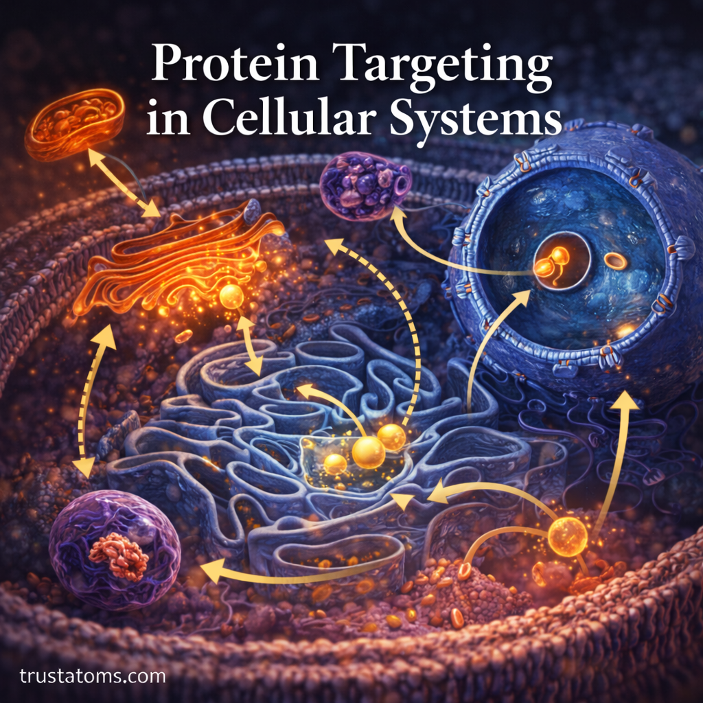
Protein targeting is the process that directs proteins to the correct locations inside or outside a cell. Cells use molecular signals and transport machinery to deliver proteins to organelles, membranes, and other destinations where they perform their functions, making protein targeting an essential concept in biology.
Most proteins in eukaryotic cells are produced by ribosomes in the cytoplasm or on the rough endoplasmic reticulum. However, their final destinations may be very different. Some enter the nucleus, others function inside mitochondria, and many travel through the endoplasmic reticulum and Golgi apparatus before reaching the cell surface or being secreted.
Cells solve this delivery problem using targeting signals that act like molecular addresses. When these signals or the machinery that recognizes them fail, proteins may accumulate in the wrong compartments, lose their function, or contribute to disease.
What Is Protein Targeting?
Protein targeting refers to the cellular mechanisms that recognize where a protein belongs and direct it toward that destination. Many proteins contain amino acid sequences or structural features that are recognized by specialized transport machinery.
Common destinations include the nucleus, mitochondria, endoplasmic reticulum, Golgi apparatus, lysosomes, plasma membrane, and extracellular space. Some proteins remain in the cytosol because they do not require transport to another compartment.
Targeting is closely connected to the broader movement of molecules described in Intracellular Protein Transport Mechanisms. The sorting and movement of proteins between cellular locations are also examined in Protein Trafficking.
Because organelles carry out different chemical activities, protein delivery supports the cooperation described in Organelle Coordination in Cellular Metabolism. The organization of these compartments is explored further in Cellular Structural Organization.
For proteins moving through the secretory pathway, membrane-bound carriers provide another level of organization. Vesicle Transport explains how these carriers move cargo between compartments and toward the cell surface.
How Targeting Signals Guide Proteins
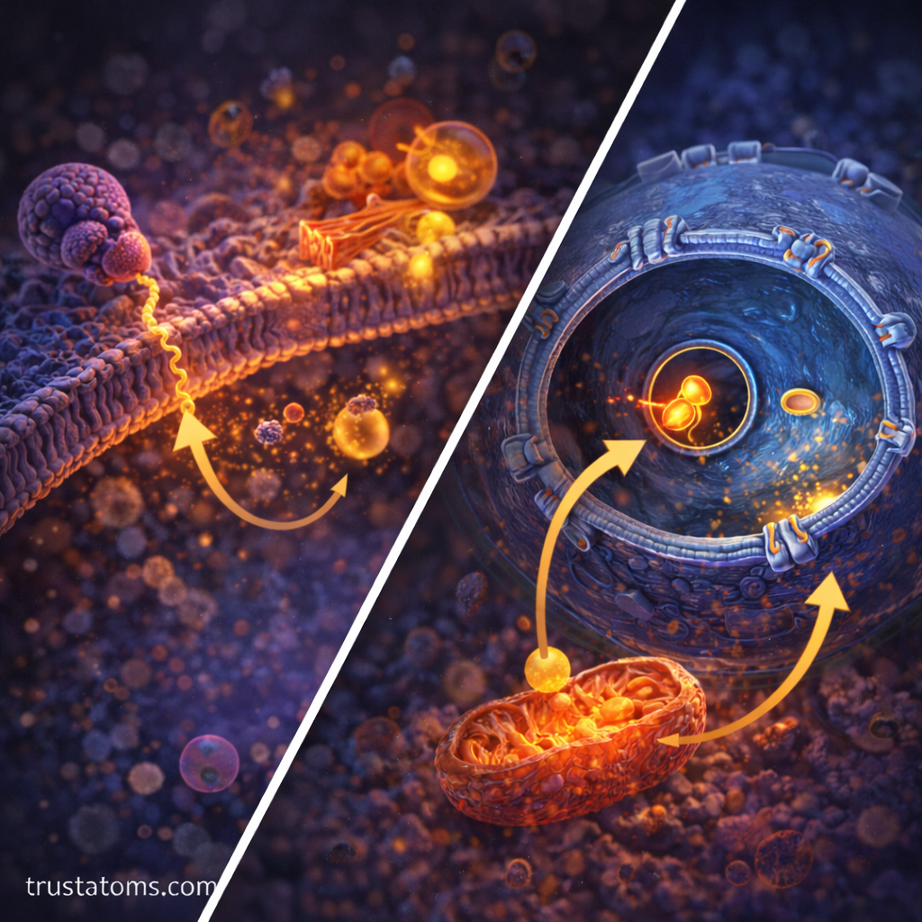
Many proteins contain targeting signals made from particular amino acid sequences. These signals are recognized by receptors or other cellular components that guide the protein toward its destination.
Signals may occur near the beginning of a protein, near its end, or within its sequence. Some targeting information depends on the protein’s three-dimensional structure rather than a single continuous sequence.
| Destination | Typical Targeting Information | Transport Machinery |
|---|---|---|
| Endoplasmic reticulum | Hydrophobic ER signal sequence | Signal recognition particle and translocon |
| Nucleus | Nuclear localization signal | Importins and nuclear pores |
| Mitochondria | Mitochondrial targeting sequence | TOM and TIM complexes |
| Peroxisomes | Peroxisomal targeting signal | Pex receptors and import machinery |
| Lysosomes | Mannose-6-phosphate tag on many soluble enzymes | Mannose-6-phosphate receptors |
These mechanisms are not interchangeable. Each destination requires specific recognition and transport processes, and some proteins use additional or alternative pathways.
How Proteins Enter the Endoplasmic Reticulum
The endoplasmic reticulum, or ER, is a major entry point for proteins destined for secretion, the plasma membrane, lysosomes, and several other compartments of the endomembrane system.
Many proteins enter the ER while they are still being synthesized. This process is called cotranslational translocation.
- Signal recognition: An ER signal sequence emerges from the ribosome and is recognized by the signal recognition particle, or SRP.
- Membrane docking: SRP helps direct the ribosome and growing protein to an SRP receptor at the ER membrane.
- Protein transfer: The ribosome associates with a protein-conducting channel called a translocon.
- Continued synthesis: As translation proceeds, the protein enters the ER lumen or becomes inserted into the ER membrane.
Inside the ER, proteins may fold, form disulfide bonds, and receive carbohydrate modifications. Some ER signal peptides are removed during translocation, while other signal sequences remain part of membrane proteins.
How the ER and Golgi Sort Proteins
After entering the ER, many proteins move toward the Golgi apparatus through membrane-bound transport carriers. This pathway allows cells to process proteins and sort them for different destinations.
Processing in the Endoplasmic Reticulum
The ER contains enzymes and molecular chaperones that assist protein folding and modification. Quality-control systems help retain or remove proteins that have not folded appropriately, reducing the chance that defective proteins continue through the secretory pathway.
Sorting in the Golgi Apparatus
The Golgi apparatus further modifies many proteins, including their attached carbohydrate groups. Sorting machinery helps direct cargo toward the plasma membrane, endosomes, lysosomes, or secretion outside the cell.
Transport carriers must recognize the correct destination membrane and fuse with it to deliver their contents. This process involves specialized proteins, including Rab GTPases and SNARE proteins.
How Proteins Enter the Nucleus
The nucleus contains DNA and the machinery involved in regulating gene expression. Many proteins required for these activities are synthesized in the cytoplasm and must enter through nuclear pore complexes.
Proteins carrying a nuclear localization signal are often recognized by transport receptors called importins. These receptors help move their cargo through nuclear pores. The Ran GTPase system contributes to the directionality of nuclear transport and cargo release.
Unlike proteins entering mitochondria, many nuclear proteins can pass through nuclear pores while remaining folded. Small molecules and some small proteins may also diffuse through pores without requiring an active import pathway.
How Mitochondria Import Proteins
Mitochondria produce much of the ATP used by eukaryotic cells. Although they contain their own DNA and protein synthesis machinery, most mitochondrial proteins are encoded by nuclear genes and synthesized on cytosolic ribosomes.
Many mitochondrial proteins contain targeting sequences recognized by transport machinery in the outer mitochondrial membrane. The TOM complex helps proteins cross this outer membrane, while TIM complexes assist the import of certain proteins across or into the inner membrane.
Protein import may require ATP, membrane potential, and molecular chaperones. Some imported proteins are subsequently processed and folded within the mitochondrion. Different mitochondrial compartments use distinct sorting pathways.
How Lysosomal Enzymes Reach Their Destination
Lysosomes contain enzymes that break down biological molecules. Many soluble lysosomal enzymes are synthesized in the ER and processed through the Golgi apparatus, where they receive a carbohydrate-based sorting marker called mannose-6-phosphate, or M6P.
M6P receptors recognize these tagged enzymes and help package them into carriers traveling toward endosomes. The enzymes are eventually delivered to lysosomes, while the receptors can be recycled for further transport.
Not all lysosomal proteins use M6P-dependent sorting. Lysosomal membrane proteins and some soluble enzymes follow alternative targeting pathways.
Real-World Example: I-Cell Disease and Protein Mistargeting
I-cell disease, also called mucolipidosis II alpha/beta, illustrates what happens when a protein-targeting system fails. This inherited disorder affects an enzyme involved in adding the mannose-6-phosphate targeting marker to many lysosomal enzymes.
Under normal conditions, the marker helps direct these enzymes toward lysosomes. In affected cells, many enzymes do not receive the appropriate targeting information and are instead secreted outside the cell.
Without sufficient amounts of certain digestive enzymes inside lysosomes, substances that would normally be broken down accumulate within cells. This contributes to severe abnormalities involving growth, skeletal development, and multiple organs.
The example demonstrates that producing a functional enzyme is not always enough. The enzyme must also reach the compartment where its activity is needed.
How Cells Detect Protein Targeting Errors
Cells have quality-control systems that help identify incorrectly folded, damaged, or misplaced proteins. These systems protect cellular compartments and limit the accumulation of potentially harmful molecules.
For example, ER-associated degradation can direct certain misfolded ER proteins toward destruction by the proteasome. Other pathways use lysosomes to break down cellular material, including proteins and damaged organelles.
Targeting defects can arise from altered signal sequences, defective transport receptors, or problems with membrane trafficking. Their consequences depend on the affected protein and its normal function.
How Scientists Study Protein Localization
Researchers combine imaging, genetic, and biochemical methods to determine where proteins travel and how targeting signals work.
- Fluorescent protein tagging: Allows researchers to visualize the location of tagged proteins in cells.
- Confocal microscopy: Helps examine protein distribution within cellular compartments.
- Signal-sequence mutations: Test whether specific amino acids are required for targeting.
- Cell fractionation: Separates cellular compartments so researchers can analyze their protein contents.
- Live-cell imaging: Makes it possible to follow some protein movements over time.
Combining methods helps distinguish a protein’s normal destination from temporary locations it passes through during transport. These approaches also help researchers investigate diseases involving abnormal protein localization.
Frequently Asked Questions
Do all proteins need targeting signals?
No. Many proteins function in the cytosol without being transported to another compartment. Others contain signals that direct them into organelles, membranes, or the secretory pathway.
What is the difference between protein targeting and protein trafficking?
Protein targeting concerns how proteins are directed to particular destinations. Protein trafficking describes their movement and sorting through cellular transport pathways. The two processes overlap because successful delivery requires both destination recognition and transport.
What happens when a protein reaches the wrong organelle?
A misplaced protein may fail to perform its normal function, interfere with cellular processes, or be recognized by quality-control machinery. The consequences range from minor changes to serious cellular dysfunction, depending on the protein involved.
Protein targeting allows cells to distribute proteins among compartments with different functions. By recognizing molecular signals and coordinating transport machinery, cells ensure that enzymes, receptors, and structural proteins reach appropriate destinations. Studying these pathways helps explain how cellular organization is maintained and how delivery errors contribute to disease.
Sources: Alberts et al., Molecular Biology of the Cell; Lodish et al., Molecular Cell Biology; National Institute of General Medical Sciences.